Product Detail
Product Namec-Myc (Phospho-T58/S62) Rabbit mAb
Clone No.SZ02-06
Host SpeciesRabbit
ClonalityMonoclonal
PurificationProA affinity purified
ApplicationsWB, ICC/IF
Species ReactivityHu, Rt
Immunogen DescSynthetic phospho-peptide corresponding to residues surrounding Thr58 and Ser62 of human c-Myc.
ConjugateUnconjugated
Other NamesAvian myelocytomatosis viral oncogene homolog antibody
bHLHe39 antibody
c Myc antibody
Class E basic helix-loop-helix protein 39 antibody
MRTL antibody
Myc antibody
Myc protein antibody
Myc proto oncogene protein antibody
Myc proto-oncogene protein antibody
myc related translation/localization regulatory factor antibody
MYC_HUMAN antibody
Myc2 antibody
MYCC antibody
Niard antibody
Nird antibody
Proto-oncogene c-Myc antibody
Transcription factor p64 antibody
v myc avian myelocytomatosis viral oncogene homolog antibody
v myc myelocytomatosis viral oncogene homolog antibody
Accession NoSwiss-Prot#:P01106
Uniprot
P01106
Gene ID
4609;
Calculated MWPredicted band size: 49 kDa
Sdspage MWObserved band size: 57 kDa
Formulation1*TBS (pH7.4), 1%BSA, 40%Glycerol. Preservative: 0.05% Sodium Azide.
StorageStore at -20˚C
Application Details
WB: 1:500-1:2000
ICC/IF: 1:50-1:200
All lanes : c-Myc (Phospho-T58/S62) Rabbit mAb at 1/1k dilutionLane 1 : C6 whole cell lysatesLane 2 : 293T whole cell lysatesLane 3 : 293T treated with 100ng/ml EGF for 20min whole cell lysatesSecondaryAll lanes : Goat Anti-Rabbit IgG H&L (HRP) at 1/20000 dilutionPredicted band size: 49 kDa Observed band size: 57 kDaExposure time: 9 seconds
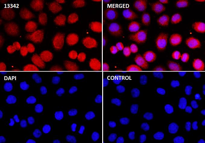
Immunocytochemistry/ Immunofluorescence c-Myc (Phospho-T58/S62) antibody (13342)
ICC/IF staining of c-Myc (Phospho-T58/S62) in HeLa cells. Cells were fixed with 4% Paraformaldehyde permeabilized with 0.1% Triton X-100.
Samples were incubated with 13342 at a working dilution of 1/100. The secondary antibody was Alexa Fluor® 647 goat anti rabbit, used at a dilution of 1/500.
The negative control is shown in bottom right hand panel - for the negative control.
Nuclei were counterstained with DAPI.
c-Myc-, N-Myc- and L-Myc-encoded proteins function in cell proliferation, differentiation and neoplastic disease. Myc proteins are nuclear proteins with relatively short half lives. Amplification of the c-Myc gene has been found in several types of human tumors including lung, breast and colon carcinomas, while the N-Myc gene has been found amplified in neuroblastomas. The L-Myc gene has been reported to be amplified and expressed at high level in human small cell lung carcinomas. The presence of three sequence motifs in the c-Myc COOH terminus, including the leucine zipper, the helix-loop-helix and a basic region provided initial evidence for a sequence-specific binding function. A basic region helix-loop-helix leucine zipper motif (bHLH-Zip) protein, designated Max, specifically associates with c-Myc, N-Myc and L-Myc proteins. The Myc-Max complex binds to DNA in a sequence-specific manner under conditions where neither Max nor Myc exhibit appreciable binding. Max can also form heterodimers with at least two additional bHLH-Zip proteins, Mad and Mxi1, and Mad-Max dimers have been shown to repress transcription through interaction with mSin3.
If you have published an article using product 13342, please notify us so that we can cite your literature.
et al,Cytoplasmic localization of SETDB1‑induced Warburg effect via c‑MYC‑LDHA axis enhances migration and invasion in breast carcinoma. In Int J Mol Med on 2024 Apr by Wenlin Yang, Yingze Wei, et al..PMID:38426579
, (2024),
PMID:
38426579

 Yes
Yes