Product Detail
Product NameIKB alpha(Phospho-S32) Rabbit mAb
Clone No.ST53-05
Host SpeciesRabbit
ClonalityMonoclonal
PurificationProA affinity purified
ApplicationsWB, ICC/IF
Species ReactivityHu
Immunogen DescSynthetic phospho-peptide corresponding to residues surrounding Ser32 of human IKB alpha.
ConjugateUnconjugated
Other NamesI kappa B alpha antibody
I-kappa-B-alpha antibody
IkappaBalpha antibody
IkB-alpha antibody
IKBA antibody
IKBA_HUMAN antibody
IKBalpha antibody
MAD 3 antibody
MAD3 antibody
Major histocompatibility complex enhancer-binding protein MAD3 antibody
NF kappa B inhibitor alpha antibody
NF-kappa-B inhibitor alpha antibody
NFKBI antibody
NFKBIA antibody
Nuclear factor of kappa light chain gene enhancer in B cells antibody
Nuclear factor of kappa light polypeptide gene enhancer in B cells inhibitor alpha antibody
Accession NoSwiss-Prot#:P25963
Uniprot
P25963
Gene ID
4792;
Calculated MWPredicted band size: 36 kDa
Sdspage MWObserved band size: 36 kDa
Formulation1*TBS (pH7.4), 1%BSA, 40%Glycerol. Preservative: 0.05% Sodium Azide.
StorageStore at -20˚C
Application Details
WB: 1:500-1:2000
ICC/IF: 1:50-1:200
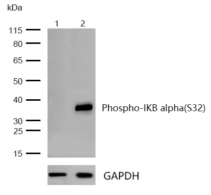
All lanes : IKB alpha(Phospho-S32) Rabbit mAb at 1/1k dilution
Lane 1 : Hela whole cell lysates
Lane 2 : Hela treated with 20ng/mL TNF-α for 10 minutes whole cell lysate
Lysates/proteins at 20 µg per lane.
Secondary
All lanes : Goat Anti-Rabbit IgG H&L (HRP) at 1/20000 dilution
Predicted band size: 36 kDa
Observed band size: 36 kDa
Exposure time: 12 seconds
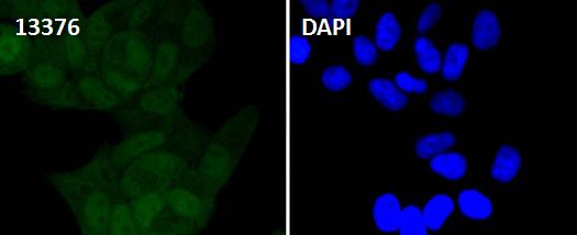
Immunocytochemistry/ Immunofluorescence IKB alpha(Phospho-S32) antibody (13376)
ICC/IF staining of IKB alpha(Phospho-S32) in HeLa cells. Cells were fixed with 4% Paraformaldehyde permeabilized with 0.1% Triton X-100.
Samples were incubated with 13376 at a working dilution of 1/100. The secondary antibody was Alexa Fluor® 488 goat anti rabbit, used at a dilution of 1/500.
Nuclei were counterstained with DAPI.
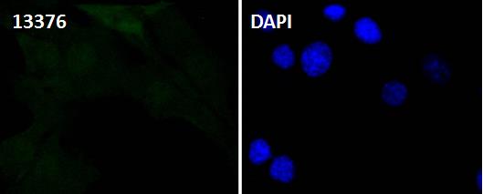
Immunocytochemistry/ Immunofluorescence IKB alpha(Phospho-S32) antibody (13376)
ICC/IF staining of IKB alpha(Phospho-S32) in NIH/3T3 cells. Cells were fixed with 4% Paraformaldehyde permeabilized with 0.1% Triton X-100.
Samples were incubated with 13376 at a working dilution of 1/100. The secondary antibody was Alexa Fluor® 488 goat anti rabbit, used at a dilution of 1/500.
Nuclei were counterstained with DAPI.
On the basis of both functional and structural considerations, members of the IκB family of proteins can be divided into four groups. The first of these groups, IκB-α, includes the avian protein pp40 and the mammalian MAD-3, both of which inhibit binding of p50-p65 NFkB complex or Rel protein to their cognate binding sites but do not inhibit the binding of p50 homodimer to κB sites, suggesting that the IκB-α family binds to the p65 subunit of p50-p65 heterocomplex through ankyrin repeats. The second member of the IκB family is represented by a protein designated IκB-β. The third group of IκB proteins is represented by IκB-γ, which is identical in sequence with the C-terminal domain of the p110 precursor of NFkB p50 and is expressed predominantly in lymphoid cells. An additional IκB family member, IκB-ε, has several phosphorylated forms and is primarily found complexed with Rel A and/or c-Rel.
If you have published an article using product 13376, please notify us so that we can cite your literature.


 Yes
Yes


