Product Detail
Product NameSHP2(Phospho-Y542) Rabbit mAb
Clone No.SN61-01
Host SpeciesRabbit
ClonalityMonoclonal
PurificationProA affinity purified
ApplicationsWB, ICC/IF
Species ReactivityHu, Ms
Immunogen DescSynthetic phospho-peptide corresponding to residues surrounding Tyr542 of human SHP2.
ConjugateUnconjugated
Other NamesBPTP3 antibody
CFC antibody
JMML antibody
METCDS antibody
MGC14433 antibody
NS1 antibody
OTTHUMP00000166107 antibody
OTTHUMP00000166108 antibody
Protein tyrosine phosphatase 2 antibody
Protein tyrosine phosphatase 2C antibody
Protein tyrosine phosphatase non receptor type 11 antibody
Protein-tyrosine phosphatase 1D antibody
Protein-tyrosine phosphatase 2C antibody
PTN11_HUMAN antibody
PTP-1D antibody
PTP-2C antibody
PTP1D antibody
PTP2C antibody
PTPN11 antibody
SAP2 antibody
SH-PTP2 antibody
SH-PTP3 antibody
SH2 domain containing protein tyrosine phosphatase 2 antibody
SHP 2 antibody
SHP-2 antibody
Shp2 antibody
SHPTP2 antibody
SHPTP3 antibody
Syp antibody
Tyrosine-protein phosphatase non-receptor type 11 antibody
Accession NoSwiss-Prot#:Q06124
Uniprot
Q06124
Gene ID
5781;
Calculated MWPredicted band size: 68 kDa
Sdspage MWObserved band size: 68 kDa
Formulation1*TBS (pH7.4), 1%BSA, 40%Glycerol. Preservative: 0.05% Sodium Azide.
StorageStore at -20˚C
Application Details
WB: 1:500-1:2000
ICC/IF: 1:50-1:200
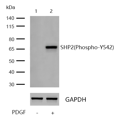
All lanes: SHP2(Phospho-Y542) Rabbit mAb at 1/1k dilution
Lane 1 : NIH/3T3 whole cell lysates
Lane 2 : NIH/3T3 treated with 40ng/mL PDGF for 40 minutes whole cell lysates
Lysates/proteins at 20 µg per lane.
Secondary
All lanes : Goat Anti-Rabbit IgG H&L (HRP) at 1/20000 dilution
Predicted band size: 68 kDa
Observed band size: 68 kDa
Exposure time: 15 seconds
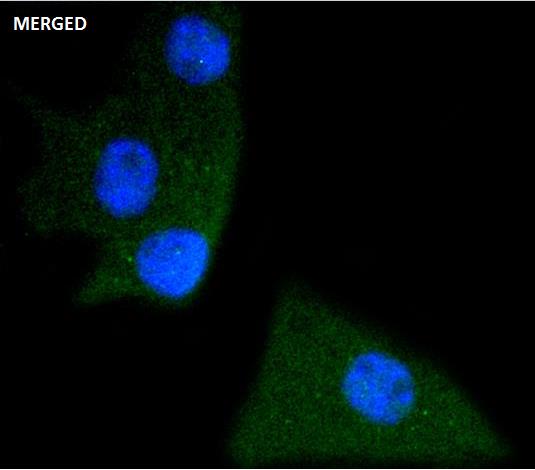
Immunocytochemistry/ Immunofluorescence SHP2 (Phospho-Y542) antibody (13393)
ICC/IF staining of SHP2(Phospho-Y542) in B6F1 cells. Cells were fixed with 4% Paraformaldehyde permeabilized with 0.1% Triton X-100.
Samples were incubated with 13393 at a working dilution of 1/100. The secondary antibody was Alexa Fluor® 488 goat anti rabbit, used at a dilution of 1/500.
Nuclei were counterstained with DAPI.
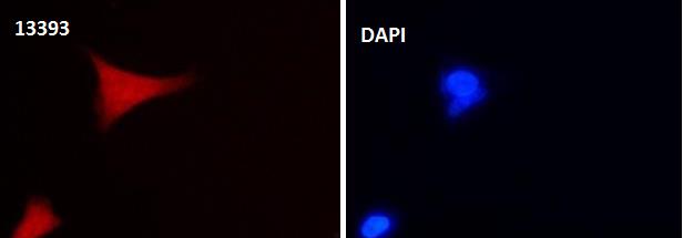
Immunocytochemistry/ Immunofluorescence SHP2 (Phospho-Y542) antibody (13393)
ICC/IF staining of SHP2(Phospho-Y542) in 293 cells. Cells were fixed with 4% Paraformaldehyde permeabilized with 0.1% Triton X-100.
Samples were incubated with 13393 at a working dilution of 1/100. The secondary antibody was Alexa Fluor® 647 goat anti rabbit, used at a dilution of 1/500.
Nuclei were counterstained with DAPI.
The steady state of protein tyrosyl phosphorylation in cells is regulated by the opposing action of tyrosine kinases and protein tyrosine phosphatases (PTPs). Several groups have independently identified a non-transmembrane PTP, designated SH-PTP1 (also known as PTP1C, HCP and SHP), which is primarily expressed in hematopoietic cells and characterized by the presence of two SH2 domains N-terminal to the PTP domain. SH2 domains generally mediate the association of regulatory molecules with specific phosphotyrosine-containing sites on autophosphorylated receptors, thereby controlling the initial interaction of receptors with these substrates. A second and much more widely expressed PTP with SH2 domains, SH-PTP2 (also designated PTP1D and Syp), has been identified. Strong sequence similarity between SH-PTP2 and the Drosophila gene corkscrew (CSW) and their similar patterns of expression suggest that SH-PTP2 is the human corkscrew homolog.
If you have published an article using product 13393, please notify us so that we can cite your literature.
et al,Grass carp (Ctenopharyngodon idellus) SHP2 suppresses IFN I expression via decreasing the phosphorylation of GSK3 beta in a non-contact manner
, (2021),
PMID:
34265416


 Yes
Yes


