Welcome to Sabbiotech!     
Email:info@signalwayantibody.com
Location: Home > Phospho Antibodies > PLK1(Phospho-T210) Rabbit mAb

PLK1(Phospho-T210) Rabbit mAb#13421

Availability: Yes
Total Amount:$
Product Detail

Product NamePLK1(Phospho-T210) Rabbit mAb

Clone No.JJ080-9

Host SpeciesRabbit

ClonalityMonoclonal

PurificationProA affinity purified

ApplicationsWB,IHC

Species ReactivityHu, Ms, Rt

Immunogen DescSynthetic phospho-peptide corresponding to residues surrounding Thr210 of human PLK1.

ConjugateUnconjugated

Other NamesCell cycle regulated protein kinase antibody
PLK 1 antibody
PLK antibody
PLK-1 antibody
plk1 antibody
PLK1_HUMAN antibody
Polo like kinase 1 antibody
Polo-like kinase 1 antibody
Serine/threonine protein kinase 13 antibody
Serine/threonine protein kinase PLK1 antibody
Serine/threonine-protein kinase 13 antibody
Serine/threonine-protein kinase PLK1 antibody
STPK 13 antibody
STPK13 antibody

Accession NoSwiss-Prot#:P53350

Uniprot P53350

Gene ID 5347;

Calculated MWPredicted band size: 68 kDa

Sdspage MWObserved band size: 68 kDa

Formulation1*TBS (pH7.4), 1%BSA, 40%Glycerol. Preservative: 0.05% Sodium Azide.

StorageStore at -20˚C

Application Details
WB: 1:500-1:2000
IHC: 1:50-1:200
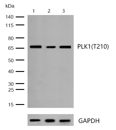
All lanes : PLK1(Phospho-T210) Rabbit mAb at 1/500 dilution

Lane 1 : Hela whole cell lysates
Lane 2 : PC12 whole cell lysates
Lane 3 : Mouse cerebellum lysates

Lysates/proteins at 20 µg per lane.

Secondary
All lanes : Goat Anti-Rabbit IgG H&L (HRP) at 1/20000 dilution

Predicted band size: 68 kDa
Observed band size: 68 kDa

Exposure time: 12 seconds
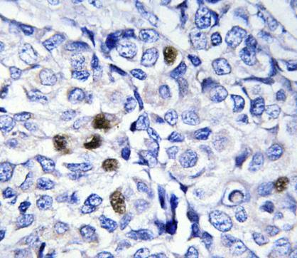
Formalin-fixed, paraffin-embedded human breast carcinoma tissue stained for PLK1 (Phospho-T210) using 13421 at 1/100 dilution in immunohistochemical analysis.
Plk (for polo-like kinase) encodes a serine/threonine kinase that is closely related to polo and CDC5, genes that are required for passage through mitosis in Drosophila and Saccharomyces, respectively. Polo and CDC5 both code for proteins that are involved in regulating the function of the mitotic spindle. Plk protein accumulates in the cell during the S and G2 phases of the cell cycle and both protein content and catalytic activity peak at the onset of mitosis, followed by a rapid reduction after mitosis. Plk expression is detectable in mitotically active tissues such as colon and placenta, as well as in tumors of various origins. It has also been suggested that Plk may serve as a marker of cell proliferation. The phosphorylation of mouse, rat and human Plk on Thr 210 enhances Plk catalytic activity.

If you have published an article using product 13421, please notify us so that we can cite your literature.

NOTE

Application

  • WBWestern Blotting
  • IHCImmunohistochemistry
  • IFImmunofluorescence
  • ICCImmunocytochemistry
  • FCFlow Cytometry
  • IPImmunoprecipitation
  • EELISA
  • DBDot Blotting
  • ChIPChromatin Immunoprecipitation
  • GICAGold Immunochromatography Assay
  • NCNegative Control

Species Reactivity

  • HuHuman
  • MsMouse
  • RtRat
  • DmDrosophila melanogaster
  • CCaenorhabditis elegans
  • MkMonkey
  • RbRabbit
  • BBovine
  • DDog
  • PPig
  • HmHamster
  • ChHmChinese Hamster
  • ChkChicken
  • ShpSheep
;