Location:
Home
>
Polyclonal Antibodies > SHP-2(Ab-542) Antibody
SHP-2(Ab-542) Antibody#21319

 Yes
Yes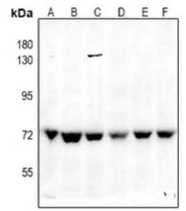
Western blot analysis of SHPTP2 expression in C6 (A), SGC7901(B), H1792 (C), SKOVCAR3 (D), A2780 (E), Hela (F) whole celllysates.
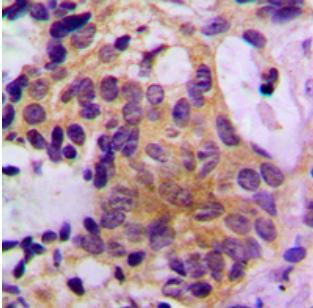
Immunohistochemical analysis of SHPTP2 staining in humanbreast cancer formalin fixed paraffin embedded tissue section.The section was pre-treated using heat mediated antigenretrieval with sodium citrate buffer (pH 6.0). The section wasthen incubated with the antibody at room temperature anddetected using an HRP conjugated compact polymer system.DAB was used as the chromogen. The section was thencounterstained with haematoxylin and mounted with DPX.
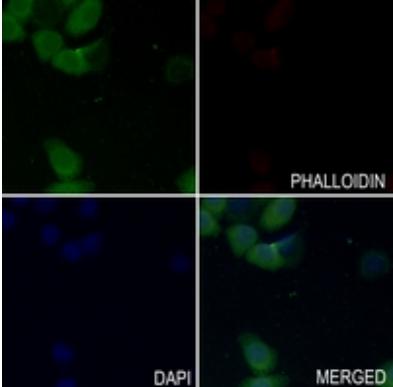
Immunofluorescent analysis of SHPTP2 staining in SGC7901cells. Formalin-fixed cells were permeabilized with 0.1% TritonX-100 in TBS for 5-10 minutes and blocked with 3% BSA-PBSfor 30 minutes at room temperature. Cells were probed withthe primary antibody in 3% BSA-PBS and incubated overnightat 4 °C in a hidified chamber. Cells were washed with PBST andincubated with a AF488-conjugated secondary antibody(green) in PBS at room temperature in the dark. Phalloidin -AF594 was used to stain Actin filaments (red). DAPI was usedto stain the cell nuclei (blue).
NOTE
Application
- WBWestern Blotting
- IHCImmunohistochemistry
- IFImmunofluorescence
- ICCImmunocytochemistry
- FCFlow Cytometry
- IPImmunoprecipitation
- EELISA
- DBDot Blotting
- ChIPChromatin Immunoprecipitation
- GICAGold Immunochromatography Assay
- NCNegative Control
Species Reactivity
- HuHuman
- MsMouse
- RtRat
- DmDrosophila melanogaster
- CCaenorhabditis elegans
- MkMonkey
- RbRabbit
- BBovine
- DDog
- PPig
- HmHamster
- ChHmChinese Hamster
- ChkChicken
- ShpSheep


