Product Detail
Product NameE-Cadherin Antibody
Clone No.A0-G11-2
Host SpeciesMouse
ClonalityMonoclonal
PurificationProA affinity purified
ApplicationsWB, ICC, IHC, FC
Species ReactivityHu,Ms
Immunogen Descrecombinant protein
ConjugateUnconjugated
Other NamesArc 1 antibody
CADH1_HUMAN antibody
Cadherin 1 antibody
cadherin 1 type 1 E-cadherin antibody
Cadherin1 antibody
CAM 120/80 antibody
CD 324 antibody
CD324 antibody
CD324 antigen antibody
cdh1 antibody
CDHE antibody
E-Cad/CTF3 antibody
E-cadherin antibody
ECAD antibody
Epithelial cadherin antibody
epithelial calcium dependant adhesion protein antibody
LCAM antibody
Liver cell adhesion molecule antibody
UVO antibody
Uvomorulin antibody
Accession NoSwiss-Prot#:P09803
Uniprot
P09803
Gene ID
12550;
Calculated MW130 kDa
Formulation1*TBS (pH7.4), 1%BSA, 40%Glycerol. Preservative: 0.05% Sodium Azide.
StorageStore at -20˚C
Application Details
WB: 1:500-1:1000
IHC: 1:200
ICC: 1:200
FC: 1:50-1:100
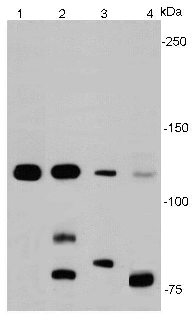
Western blot analysis of E-cadherin on different cell lysates using anti- E-cadherin antibody at 1/1000 dilution. Positive control:
Lane 1: A431
Lane 2: SW480
Lane 3: MCF-7
Lane 4: Hela
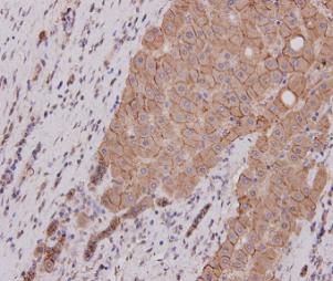
Immunohistochemical analysis of paraffin-embedded human liver carcinoma tissue using E-Cadherin antibody. Counter stained with hematoxylin.
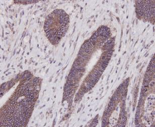
Immunohistochemical analysis of paraffin-embedded human colon carcinoma tissue using E-Cadherin antibody. Counter stained with hematoxylin.
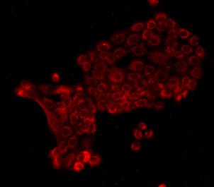
ICC staining E-Cadherin in A431 cells (red). Cells were fixed in paraformaldehyde, permeabilised with 0.25% Triton X100/PBS.
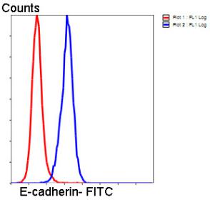
Flow cytometric analysis of HeLa cells with E-Cadherin antibody at 1/50 dilution (blue) compared with an unlabelled control (cells without incubation with primary antibody; red). Goat anti mouse IgG (FITC) was used as the secondary antibody
E-cadherin (epithelial) is the most well-studied member of the cadherin family. It consists of 5 cadherin repeats (EC1 ~ EC5) in the extracellular domain, one transmembrane domain, and an intracellular domain that binds p120-catenin and beta-catenin. The intracellular domain contains a highly-phosphorylated region vital to beta-catenin binding and, therefore, to E-cadherin function. Loss of E-cadherin function or expression has been implicated in cancer progression and metastasis. E-cadherin downregulation decreases the strength of cellular adhesion within a tissue, resulting in an increase in cellular motility. This in turn may allow cancer cells to cross the basement membrane and invade surrounding tissues. E-cadherin is also used by pathologists to diagnose different kinds of breast cancer.
If you have published an article using product 48355, please notify us so that we can cite your literature.
et al,ADAMTS13 attenuates renal fibrosis by suppressing thrombospondin 1 mediated TGF-β1/Smad3 activation.
, (2025),
PMID:
39929281


 Yes
Yes


