Product Detail
Product NamePKN1 Antibody
Clone No.B1-10H
Host SpeciesMouse
ClonalityMonoclonal
PurificationProA affinity purified
ApplicationsWB, IHC, ICC, FC
Species ReactivityHu
Immunogen DescRecombinant protein
ConjugateUnconjugated
Other NamesDBK antibody
PAK 1 antibody
PAK-1 antibody
PAK1 antibody
PKC1 antibody
PKN ALPHA antibody
PKN antibody
Pkn1 antibody
PKN1_HUMAN antibody
PRK1 antibody
PRKCL1 antibody
Protease activated kinase 1 antibody
Protease-activated kinase 1 antibody
Protein kinase C like 1 antibody
Protein kinase C like PKN antibody
Protein kinase C related kinase 1 antibody
Protein kinase C-like 1 antibody
Protein kinase C-like PKN antibody
Protein kinase N1 antibody
Protein kinase PKN alpha antibody
Protein kinase PKN-alpha antibody
Protein-kinase C-related kinase 1 antibody
Serine threonine kinase N antibody
Serine threonine protein kinase N antibody
Serine-threonine protein kinase N antibody
Serine/threonine protein kinase N1 antibody
Serine/threonine-protein kinase N1 antibody
Accession NoSwiss-Prot#:Q16512
Uniprot
Q16512
Gene ID
5585;
Calculated MW104 kDa
Formulation1*TBS (pH7.4), 1%BSA, 40%Glycerol. Preservative: 0.05% Sodium Azide.
StorageStore at -20˚C
Application Details
WB: 1:500-1:2,000
IHC: 1:50-1:200
ICC: 1:50-1:200
FC: 1:50-1:100
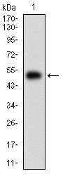
Western blot analysis of PKN1 on human PKN1 recombinant protein using anti-PKN1 antibody at 1/1,000 dilution.
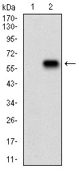
Western blot analysis of PKN1 on HEK293 (1) and PKN1-hIgGFc transfected HEK293 (2) cell lysate using anti-PKN1 antibody at 1/1,000 dilution.
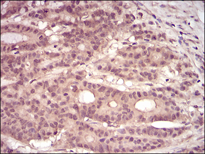
Immunohistochemical analysis of paraffin-embedded human colon cancer tissue using anti-PKN1 antibody. Counter stained with hematoxylin.
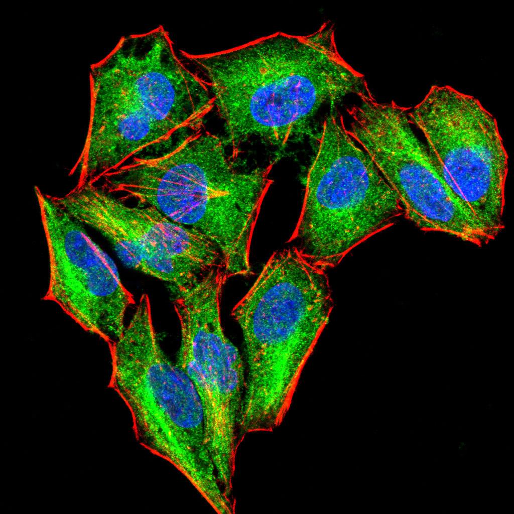
ICC staining PKN1 (green) and Actin filaments (red) in Hela cells. The nuclear counter stain is DAPI (blue). Cells were fixed in paraformaldehyde, permeabilised with 0.25% Triton X100/PBS.
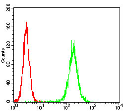
Flow cytometric analysis of Hela cells with PKN1 antibody at 1/100 dilution (green) compared with an unlabelled control (cells without incubation with primary antibody; red).
Rho, the Ras-related small GTPase, is responsible for the regulation of actin-based cytoskeletal structures including stress fibers, focal adhesions and the contractile ring apparatus. Rho proteins act as molecular switches which are able to turn cytokinesis on and off. Although little is know about signaling downstream of Rho, several proteins have been implicated as Rho effectors. Protein kinase N (PKN) is a fatty acid-activated serine/threonine kinase whose catalytic domain exhibits homology with that of the PKC family. PKN associates with Rho via its amino terminus, is activated in a GTP-dependent manner and phosphorylates the head-rod domain of neurofilament protein. A second protein, rhophilin, exhibits 40% sequence identity with the amino terminal Rho binding domain. The enzymatic activity of rhophilin has not been demonstrated and it is possible that it acts through the recruitment of cytoskeletal components that initiate a kinase signaling cascade. Citron interacts specifically with active Rho and Rac1 but not Cdc42. Citron exhibits a distinctive protein organization and little homology with the Rho binding domains of PKN and rhophilin.
If you have published an article using product 48392, please notify us so that we can cite your literature.

 Yes
Yes