Product Detail
Product NamePBK Antibody
Clone No.E11-C8
Host SpeciesMouse
ClonalityMonoclonal
PurificationProA affinity purified
ApplicationsWB,ICC,IHC,FC
Species ReactivityHu
Immunogen DescRecombinant protein
ConjugateUnconjugated
Other NamesCancer/testis antigen 84 antibody
CT84 antibody
Epididymis luminal protein 164 antibody
FLJ14385 antibody
HEL164 antibody
Lymphokine activated killer T cell originated protein kinase antibody
Lymphokine-activated killer T-cell-originated protein kinase antibody
MAPKK like protein kinase antibody
MAPKK-like protein kinase antibody
Nori 3 antibody
Nori-3 antibody
Nori3 antibody
PBK antibody
PDZ binding kinase antibody
PDZ-binding kinase antibody
Serine/threonine protein kinase antibody
Spermatogenesis related protein kinase antibody
Spermatogenesis-related protein kinase antibody
SPK antibody
T LAK cell originated protein kinase antibody
T-LAK cell-originated protein kinase antibody
TOPK antibody
TOPK_HUMAN antibody
Accession NoSwiss-Prot#:Q96KB5
Uniprot
Q96KB5
Gene ID
55872;
Calculated MW36 kDa
Formulation1*TBS (pH7.4), 1%BSA, Preservative: 0.05% Sodium Azide.
StorageStore at -20˚C
Application Details
WB: 1:500
IHC: 1:100-1:500
ICC: 1:100-1:500
FC: 1:100-1:200
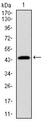
Western blot analysis of PBK on human PBK recombinant protein using anti-PBK antibody at 1/1,000 dilution.
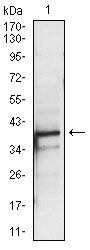
Western blot analysis of PBK on A431 cell lysate using anti-PBK antibody at 1/1,000 dilution.
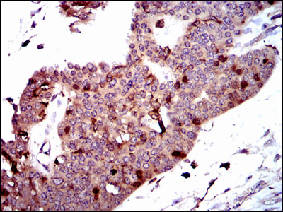
Immunohistochemical analysis of paraffin-embedded human ovarian cancer tissue using anti-PBK antibody. Counter stained with hematoxylin.
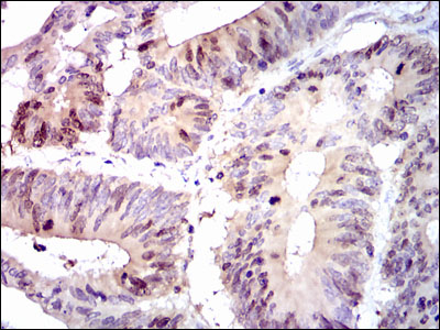
Immunohistochemical analysis of paraffin-embedded human colon cancer tissue using anti-PBK antibody. Counter stained with hematoxylin.
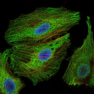
ICC staining PBK (green) and actin filaments (red) in Hela cells. The nuclear counter stain is DAPI (blue). Cells were fixed in paraformaldehyde, permeabilised with 0.25% Triton X100/PBS.
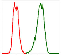
Flow cytometric analysis of Hela cells with PBK antibody at 1/100 dilution (green) compared with an unlabelled control (cells without incubation with primary antibody; red).
Protein kinases comprise a large group of encoded factors that regulate cellular processes by catalyzing the transfer of a phosphate group to a hydroxyl acceptor in serine, threonine or tyrosine residues. Kinases are capable of influencing the oncogenic potential of cell sytems at the level of oncoprotein or tumor suppressor protein phosphorylation states. Human PDZ-binding kinase, known as PBK, is a 322 amino acid, T/SXV motif-containing serine/threonine kinase that is abuntant in placenta and absent from adult brain tissue. A PDZ domain in the tumor suppressor protein Dlg can coordinate with the T/SXV motif of PBK. The cell cycle checkpoint kinase Cdc2/cyclin B is an upstream effector of PBK that can phosphorylate and activate PBK. Active PBK may associate with PDZ-containing proteins and influence cell cycle control or cellular proliferation.
If you have published an article using product 48483, please notify us so that we can cite your literature.

 Yes
Yes