Product Detail
Product NameBmi1 Antibody
Clone No.B3-G5
Host SpeciesMouse
ClonalityMonoclonal
PurificationProA affinity purified
ApplicationsWB, ICC, IHC, FC
Species ReactivityHu, Ms, Rt
Immunogen DescRecombinant protein within human Bmi1 full sequence.
ConjugateUnconjugated
Other NamesB lymphoma Mo MLV insertion region (mouse) antibody
B lymphoma Mo MLV insertion region 1 homolog antibody
Bmi 1 antibody
BMI1 antibody
BMI1 polycomb ring finger oncogene antibody
BMI1_HUMAN antibody
Flvi 2/bmi 1 antibody
FLVI2/BMI1 antibody
MGC12685 antibody
Murine leukemia viral (bmi 1) oncogene homolog antibody
Oncogene BMI 1 antibody
PCGF 4 antibody
PCGF4 antibody
Polycomb complex protein BMI 1 antibody
Polycomb complex protein BMI-1 antibody
Polycomb group protein Bmi1 antibody
Polycomb group ring finger 4 antibody
Polycomb group RING finger protein 4 antibody
RING finger protein 51 antibody
RNF 51 antibody
RNF51 antibody
Accession NoSwiss-Prot#:P35226
Uniprot
P35226
Gene ID
100532731;648;
Calculated MW37kDa
Formulation1*TBS (pH7.4), 1%BSA, 40%Glycerol. Preservative: 0.05% Sodium Azide.
StorageStore at -20˚C
Application Details
WB: 1:1,000
IHC: 1:200
ICC: 1:200
FC: 1:100-1:200
Western blot analysis of Bmi1 on different lysates using anti-Bmi1 antibody at 1/1,000 dilution. Positive control: Lane 1: 293T Lane 2: Jurkat Lane 3: Hela Lane 4: MCF-7 Lane 5: HepG2 Lane 6: NIH/3T3 Lane 7: PC12 Lane 8: Mouse kidney Lane 9: Human kidney Lane 10: K562 Lane 11: Human brain
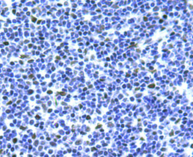
Immunohistochemical analysis of paraffin-embedded human tonsil tissue using anti-Bmi1 antibody. Counter stained with hematoxylin.
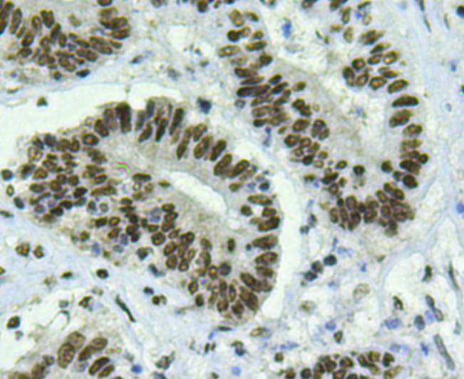
Immunohistochemical analysis of paraffin-embedded human colon cancer tissue using anti-Bmi1 antibody. Counter stained with hematoxylin.
Immunohistochemical analysis of paraffin-embedded human breast cancer tissue using anti-BMI1 antibody. Counter stained with hematoxylin.
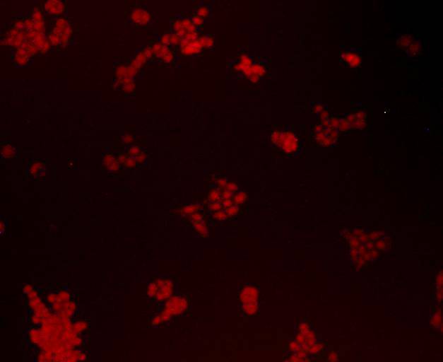
ICC staining Bmi1 in A549 cells (red). Cells were fixed in paraformaldehyde, permeabilised with 0.25% Triton X100/PBS.
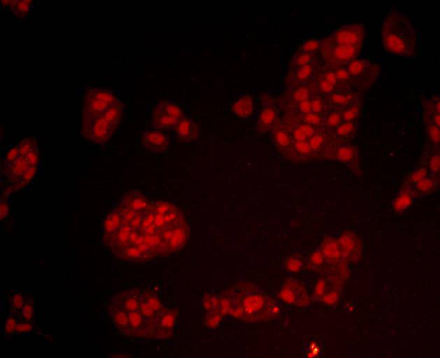
ICC staining Bmi1 in Lovo cells (red). Cells were fixed in paraformaldehyde, permeabilised with 0.25% Triton X100/PBS.
ICC staining Bmi1 in Hela cells (red). Cells were fixed in paraformaldehyde, permeabilised with 0.25% Triton X100/PBS.
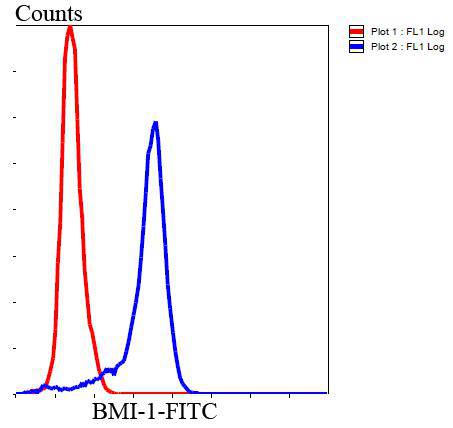
Flow cytometric analysis of Hela cells with BMI1 antibody at 1/100 dilution (blue) compared with an unlabelled control (cells without incubation with primary antibody; red). Goat anti mouse IgG (FITC) was used as the secondary antibody.
The Bmi-1 was identified initially as an oncogene that cooperates with c-myc in the generation of B-cell lymphoma. It contributes to the maintenance of cell identity, stem cell self-renewal, cell cycle regulation, and oncogenesis by maintaining the silenced state of genes that promote cell lineage specification, cell death, and cell-cycle arrest.
If you have published an article using product 48484, please notify us so that we can cite your literature.




 Yes
Yes


