Welcome to Sabbiotech!     
Email:info@signalwayantibody.com
Location: Home > Loading Controls > Beta-Actin Antibody

Beta-Actin Antibody#48485

Availability: Yes
Total Amount:$
Product Detail

Product NameBeta-Actin Antibody

Clone No.MR4561

Host SpeciesMouse

ClonalityMonoclonal

PurificationProA affinity purified

ApplicationsELISA,WB,IF,IHC,FC,IP

Species ReactivityHu, Ms, Rt

Immunogen DescSynthetic peptide (KLH-coupled) within human Beta-actin N terminal.

ConjugateUnconjugated

Other NamesA26C1A antibody
A26C1B antibody
ACTB antibody
ACTB_HUMAN antibody
Actin beta antibody
Actin cytoplasmic 1 antibody
Actin, cytoplasmic 1, N-terminally processed antibody
Actx antibody
b actin antibody
Beta cytoskeletal actin antibody
Beta-actin antibody
BRWS1 antibody
E430023M04Rik antibody
MGC128179 antibody
PS1TP5 binding protein 1 antibody
PS1TP5BP1 antibody

Accession NoSwiss-Prot#:P60709

Uniprot P60709

Gene ID 60;

Calculated MW42kDa

Formulation1*TBS (pH7.4), 1%BSA, 40%Glycerol. Preservative: 0.05% Sodium Azide.

StorageStore at -20˚C

Application Details
WB: 1:10,000
IHC: 1:100-1:200
ICC: 1:100-1:200

FC: 1:100-1:200
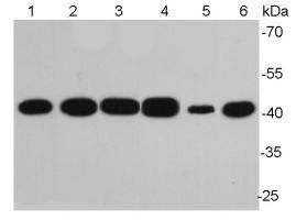
Western blot analysis of Beta-Western blot analysis of Beta-actin on different lysates using anti-Beta-actin antibody at 1/10,000 dilution.

Positive control:

Lane 1: NIH/3T3

Lane 2: PC12

Lane 3: MCF-7

Lane 4: HepG2

Lane 5: Hela

Lane 6: Mouse lung

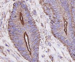
Immunohistochemical analysis of paraffin-embedded human colon carcinoma tissue using anti-Beta-actin antibody. Counter stained with hematoxylin.
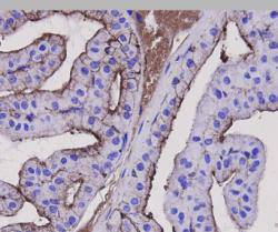
Immunohistochemical analysis of paraffin-embedded mouse prostate tissue using anti-Beta-actin antibody. Counter stained with hematoxylin.
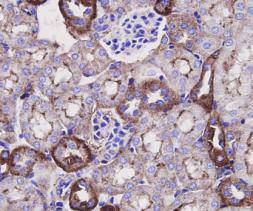
Immunohistochemical analysis of paraffin-embedded mouse kidney tissue using anti-Beta-actin antibody. Counter stained with hematoxylin.
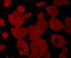
ICC staining of Beta-actin in Hela cells (red). Cells were fixed in paraformaldehyde, permeabilised with 0.25% Triton X100/PBS.
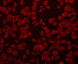
ICC staining of Beta-actin in A549 cells (red). Cells were fixed in paraformaldehyde, permeabilised with 0.25% Triton X100/PBS.
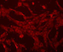
ICC staining of Beta-actin in NIH-3T3 cells (red). Cells were fixed in paraformaldehyde, permeabilised with 0.25% Triton X100/PBS.
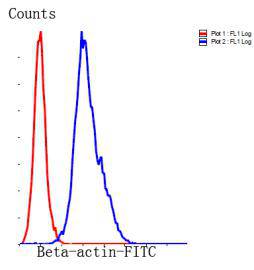
Flow cytometric analysis of Hela?cells with Beta-actin antibody at 1/100 dilution (blue) compared with an unlabelled control (cells without incubation with primary antibody; red). Goat anti mouse IgG (FITC) was used as the secondary antibody.
Beta actin also know as Cytoplasmic Actin is a 42 kDa, highly conserved protein, ubiquitously expressed in all eukaryotic cells. They comprise, along with microtubules, a major component of the cytoskeleton. β-Actin is a relatively stable cytoskeletal protein normally at a constant level in cells, regardless of experimental treatment or technical procedure. For this reason, measurement of β -Actin is generally used as an internal control for experimental error.

If you have published an article using product 48485, please notify us so that we can cite your literature.

NOTE

Application

  • WBWestern Blotting
  • IHCImmunohistochemistry
  • IFImmunofluorescence
  • ICCImmunocytochemistry
  • FCFlow Cytometry
  • IPImmunoprecipitation
  • EELISA
  • DBDot Blotting
  • ChIPChromatin Immunoprecipitation
  • GICAGold Immunochromatography Assay
  • NCNegative Control

Species Reactivity

  • HuHuman
  • MsMouse
  • RtRat
  • DmDrosophila melanogaster
  • CCaenorhabditis elegans
  • MkMonkey
  • RbRabbit
  • BBovine
  • DDog
  • PPig
  • HmHamster
  • ChHmChinese Hamster
  • ChkChicken
  • ShpSheep
;