Welcome to Sabbiotech!     
Email:info@signalwayantibody.com
Location: Home > Recombinant Rabbit Monoclonal Antibodies > LAMP2a Rabbit mAb

LAMP2a Rabbit mAb#48611

Availability: Yes
Total Amount:$
Product Detail

Product NameLAMP2a Rabbit mAb

Clone No.SA46-01

Host SpeciesRecombinant Rabbit

Clonality Monoclonal

PurificationProA affinity purified

ApplicationsWB, IHC, IP

Species ReactivityHu, Ms, Rt

Immunogen Descrecombinant protein

ConjugateUnconjugated

Other NamesCD107 antigen-like family member B antibody
CD107b antibody
LAMP 2 antibody
Lamp 2a antibody
LAMP-2 antibody
LAMP2 antibody
LAMP2_HUMAN antibody
Lysosome-associated membrane glycoprotein 2 antibody
Lysosome-associated membrane protein 2 antibody

Accession NoSwiss-Prot#:P13473

Uniprot P13473

Gene ID 3920;

Calculated MW120 kDa

Formulation1*TBS (pH7.4), 1%BSA, 40%Glycerol. Preservative: 0.05% Sodium Azide.

StorageStore at -20˚C

Application Details
WB: 1:1,000-5,000
IHC: 1:50-1:200


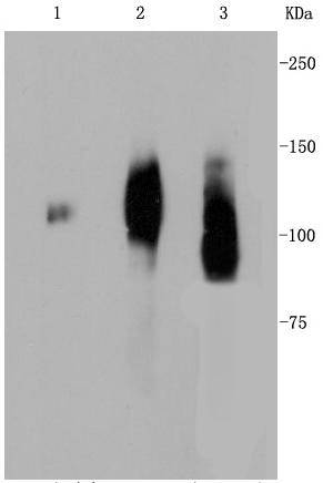
Western blot analysis of LAMP2a on different lysates using anti-LAMP2a antibody at 1/1,000 dilution. Positive control:
Lane 1: Human placenta
Lane 2: JAR
Lane 3: Human liver
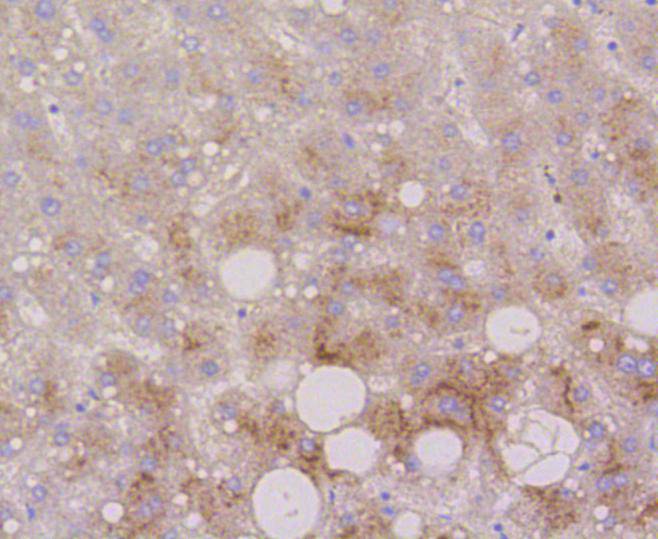
Immunohistochemical analysis of paraffin-embedded human liver tissue using anti-LAMP2a antibody. Counter stained with hematoxylin.
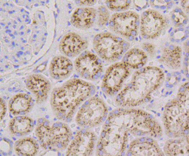
Immunohistochemical analysis of paraffin-embedded human kidney tissue using anti-LAMP2a antibody. Counter stained with hematoxylin.
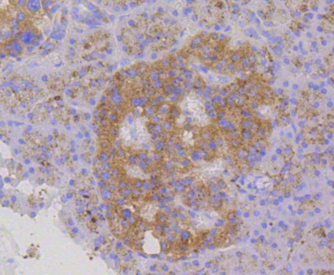
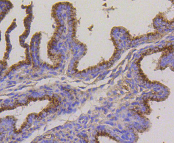
Immunohistochemical analysis of paraffin-embedded human pancreas tissue using anti-LAMP2a antibody. Counter stained with hematoxylin.
Immunohistochemical analysis of paraffin-embedded mouse kidney tissue using anti-LAMP2a antibody. Counter stained with hematoxylin.
Immunohistochemical analysis of paraffin-embedded mouse placenta tissue using anti-LAMP2a antibody. Counter stained with hematoxylin.
Immunohistochemical analysis of paraffin-embedded mouse brain tissue using anti-LAMP2a antibody. Counter stained with hematoxylin.
Lysosome-associated membrane proteins (LAMP) are glycosylated type I membrane proteins that play a role in the biogenesis of the pigment melanin. LAMP-1 (also designated CD107A) and LAMP-2 (also designated CD107B) are involved in a variety of functions, including cellular adhesion, and are thought to participate in the process of tumor invasion and metastasis. Newly synthesized LAMP-1 and LAMP-2 proteins are sorted at the trans Golgi network and are transported intracellularly via a pathway that is distinct from the clathrin-coated vesicles used for the mannose-6 phosphate receptor. LAMP-1 is expressed on the surface of thrombin-activated but not resting platelets, and it is thought to be involved in the adhesive, prothrombic properties of these cells. Both LAMP-1 and LAMP-2 are involved in maintaining lysosome acidity and protecting the lysosomal membranes from autodigestion, and their expression is increased in patients with lysosomal storage disorders.

If you have published an article using product 48611, please notify us so that we can cite your literature.

NOTE

Application

  • WBWestern Blotting
  • IHCImmunohistochemistry
  • IFImmunofluorescence
  • ICCImmunocytochemistry
  • FCFlow Cytometry
  • IPImmunoprecipitation
  • EELISA
  • DBDot Blotting
  • ChIPChromatin Immunoprecipitation
  • GICAGold Immunochromatography Assay
  • NCNegative Control

Species Reactivity

  • HuHuman
  • MsMouse
  • RtRat
  • DmDrosophila melanogaster
  • CCaenorhabditis elegans
  • MkMonkey
  • RbRabbit
  • BBovine
  • DDog
  • PPig
  • HmHamster
  • ChHmChinese Hamster
  • ChkChicken
  • ShpSheep
;