Welcome to Sabbiotech!     
Email:info@signalwayantibody.com
Location: Home > Recombinant Rabbit Monoclonal Antibodies > ERK1/2 Rabbit mAb

ERK1/2 Rabbit mAb#48616

Availability: Yes
Total Amount:$
Product Detail

Product NameERK1/2 Rabbit mAb

Clone No.SA43-03

Host SpeciesRecombinant Rabbit

ClonalityMonoclonal

PurificationProA affinity purified

ApplicationsWB, ICC/IF, IP, FC

Species ReactivityHu, Ms, Rt, zebrafish

Immunogen Descrecombinant protein

ConjugateUnconjugated

Other NamesERK 1 antibody
ERK 2 antibody
ERK-2 antibody
ERK1 antibody
erk1/2 antibody
ERK2 antibody
ERT1 antibody
ERT2 antibody
Extracellular signal regulated kinase 1 antibody
Extracellular signal-regulated kinase 2 antibody
MAP kinase 1 antibody
MAP kinase 2 antibody
MAP kinase isoform p42 antibody
MAP kinase isoform p44 antibody
MAPK 1 antibody
MAPK 2 antibody
MAPK 3 antibody
Mapk1 antibody
MAPK2 antibody
MAPK3 antibody
Mitogen-activated protein kinase 1 antibody
Mitogen-activated protein kinase 2 antibody
MK01_HUMAN antibody
p38 antibody
p40 antibody
p41 antibody
p42-MAPK antibody
PRKM 2 antibody

Accession NoSwiss-Prot#:P27361

Uniprot P27361

Gene ID 5595;

Calculated MW42/44 kDa

Formulation1*TBS (pH7.4), 1%BSA, 40%Glycerol. Preservative: 0.05% Sodium Azide.

StorageStore at -20˚C

Application Details
WB: 1:1,000-5,000

ICC: 1:50-1:200

FC: 1:50-1:100
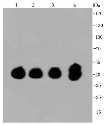
Western blot analysis of ERK1/2 on different cell lysates using anti-ERK1/2 antibody at 1/1,000 dilution. Positive control:
Lane 1: Hela
Lane 2: SW480
Lane 3: HCT116
Lane 4: PC12
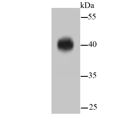
Western blot analysis of ERK1/2 on hybrid fish (crucian-carp) brain tissue lysate using anti-ERK1/2 antibody at 1/500 dilution.

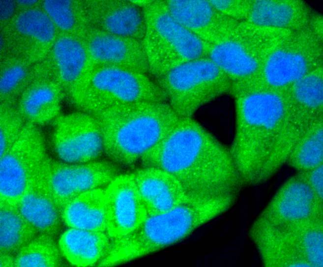
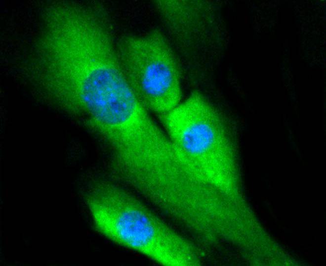
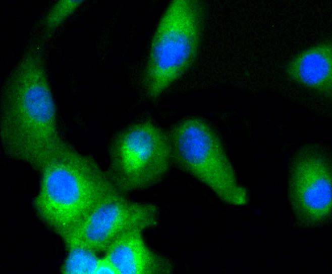
ICC staining ERK1/2 in Hela cells (green). The nuclear counter stain is DAPI (blue). Cells were fixed in paraformaldehyde, permeabilised with 0.25% Triton X100/PBS.
ICC staining ERK1/2 in MCF-7 cells (green). The nuclear counter stain is DAPI (blue). Cells were fixed in paraformaldehyde, permeabilised with 0.25% Triton X100/PBS.
ICC staining ERK1/2 in NIH/3T3 cells (green). Cells were fixed in paraformaldehyde, permeabilised with 0.25% Triton X100/PBS.
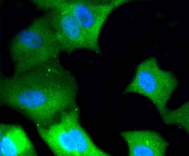
ICC staining ERK1/2 in A549 cells (green). Cells were fixed in paraformaldehyde, permeabilised with 0.25% Triton X100/PBS.
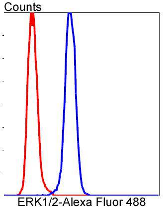
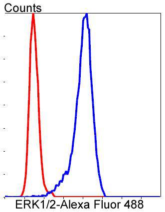
Flow cytometric analysis of Hela cells with ERK1/2 antibody at 1/50 dilution (blue) compared with an unlabelled control (cells without incubation with primary antibody; red). Alexa Fluor 488-conjugated goat anti rabbit IgG was used as the secondary antibody.
Flow cytometric analysis of SH-SY-5Y cells with ERK1/2 antibody at 1/50 dilution (blue) compared with an unlabelled control (cells without incubation with primary antibody; red). Alexa Fluor 488-conjugated goat anti rabbit IgG was used as the secondary antibody.
Mitogen-activated protein kinase (MAPK) signaling pathways involve two closely related MAP kinases, known as extracellular-signal-related kinase 1 (ERK 1, p44) and 2 (ERK 2, p42). Growth factors, steroid hormones, G protein-coupled receptor ligands and neurotransmitters can initiate MAPK signaling pathways. Activation of ERK 1 and ERK 2 requires phosphorylation by upstream kinases such as MAP kinasekinase (MEK), MEK kinase and Raf-1. ERK 1 and ERK 2 phosphorylation can occur at specific tyrosine and threonine sites mapping within consensus motifs that include the threonine-glutamate-tyrosine motif. ERK activation leads to dimerization with other ERKs and subsequent localization to the nucleus. Active ERK dimers phosphorylate serine and threonine residues on nuclear proteins and influence a host of responses that include proliferation, differentiation, transcription regulation and development. The human ERK 1 gene maps to chromosome 16p11.2 and encodes a 379 amino acid protein that shares 83% sequence identity to ERK 2. The human ERK2 gene maps to chromosome 22q11.21 and encodes a 360-amino acid protein.

If you have published an article using product 48616, please notify us so that we can cite your literature.

et al,Apoptosis-inducing activity of synthetic hydrocarbon-stapled peptides in H358 cancer cells expressing KRASG12C. In Acta Pharm Sin B on 2021 Sep by Cuicui Li, Ni Zhao,et al..PMID:34589388 , (2021),
PMID: 34589388
et al,The Effect of Clostridium butyricum-Derived Lipoteichoic Acid on Lipopolysaccharide-Stimulated Porcine Intestinal Epithelial Cells. , (2025),
PMID: 39749788

NOTE

Application

  • WBWestern Blotting
  • IHCImmunohistochemistry
  • IFImmunofluorescence
  • ICCImmunocytochemistry
  • FCFlow Cytometry
  • IPImmunoprecipitation
  • EELISA
  • DBDot Blotting
  • ChIPChromatin Immunoprecipitation
  • GICAGold Immunochromatography Assay
  • NCNegative Control

Species Reactivity

  • HuHuman
  • MsMouse
  • RtRat
  • DmDrosophila melanogaster
  • CCaenorhabditis elegans
  • MkMonkey
  • RbRabbit
  • BBovine
  • DDog
  • PPig
  • HmHamster
  • ChHmChinese Hamster
  • ChkChicken
  • ShpSheep
;