Product Detail
Product NameIFNAR1 Rabbit mAb
Clone No.SR45-08
Host SpeciesRecombinant Rabbit
Clonality Monoclonal
PurificationProA affinity purified
ApplicationsWB, IP, IHC, FC
Species ReactivityHu, Ms, Rt
Immunogen Descrecombinant protein
ConjugateUnconjugated
Other NamesAlpha type antiviral protein antibody
AVP antibody
Beta type antiviral protein antibody
CRF2-1 antibody
Cytokine receptor class-II member 1 antibody
Cytokine receptor family 2 member 1 antibody
IFN alpha REC antibody
IFN alpha receptor antibody
IFN alpha/beta Receptor alpha antibody
IFN beta receptor antibody
IFN-alpha/beta receptor 1 antibody
IFN-R-1 antibody
IFNAR antibody
Ifnar1 antibody
IFNBR antibody
IFRC antibody
INAR1_HUMAN antibody
Interferon (alpha beta and omega) receptor 1 antibody
Interferon alpha/beta receptor 1 antibody
Interferon alpha/beta receptor alpha chain antibody
Interferon beta receptor 1 antibody
Type I interferon receptor 1 antibody
Accession NoSwiss-Prot#:P17181
Uniprot
P17181
Gene ID
3454;
Calculated MW90/130 kDa
Formulation1*TBS (pH7.4), 1%BSA, 40%Glycerol. Preservative: 0.05% Sodium Azide.
StorageStore at -20˚C
Application Details
WB: 1:1,000-1:2,000
IHC:1:50-1:200
FC: 1:50-1:100
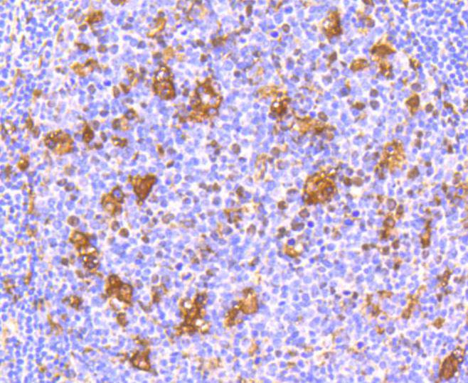
Immunohistochemical analysis of paraffin-embedded human tonsil tissue using anti-IFNAR1 antibody. Counter stained with hematoxylin.
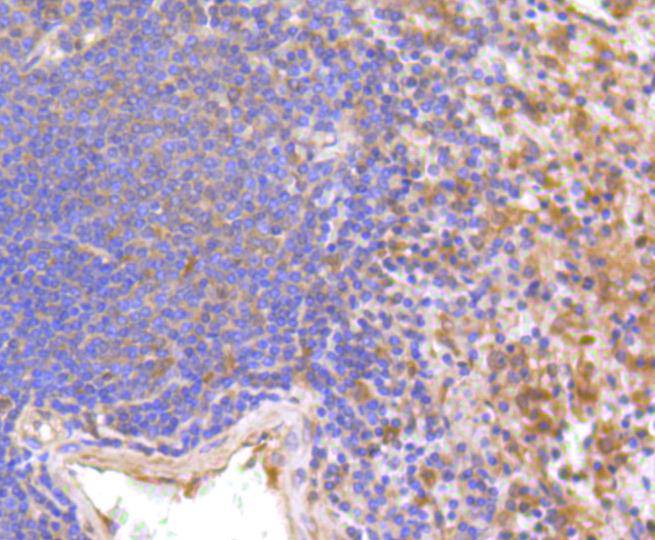
Immunohistochemical analysis of paraffin-embedded human spleen tissue using anti-IFNAR1 antibody. Counter stained with hematoxylin.
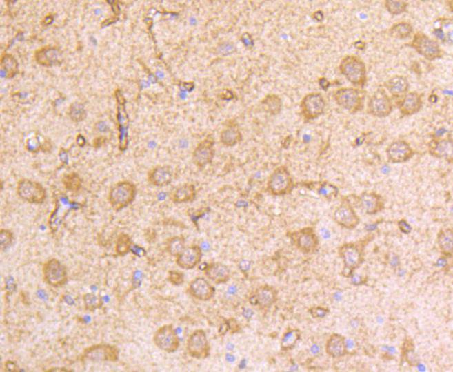
Immunohistochemical analysis of paraffin-embedded rat brain tissue using anti-IFNAR1 antibody. Counter stained with hematoxylin.
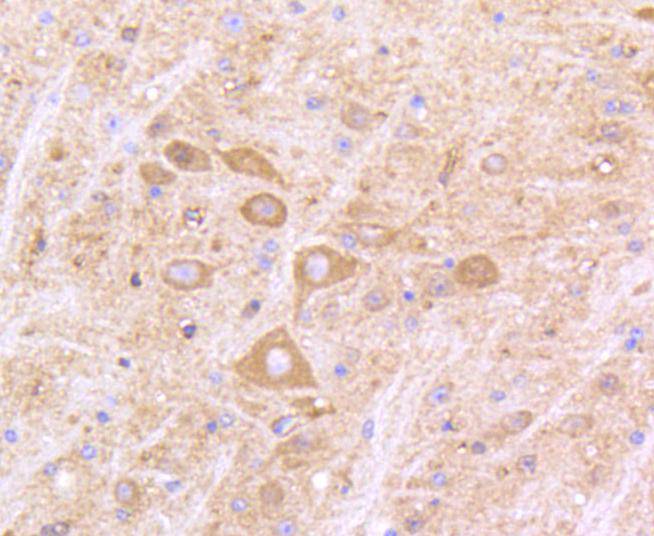
Immunohistochemical analysis of paraffin-embedded mouse brain tissue using anti-IFNAR1 antibody. Counter stained with hematoxylin.
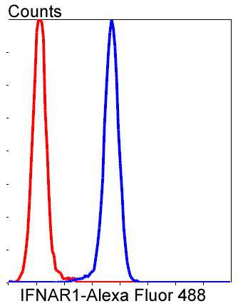
Flow cytometric analysis of Jurkat cells with IFNAR1 antibody at 1/50 dilution (blue) compared with an unlabelled control (cells without incubation with primary antibody; red). Alexa Fluor 488-conjugated goat anti rabbit IgG was used as the secondary antibody.
The type I interferons (IFNs), α and β, are a group of structurally and functionally related proteins that are induced by either viruses or double stranded RNA and defined by their ability to confer an antiviral state in cells. The α and β IFNs appear to compete with one another for binding to a common cell surface receptor, while immune IFN (IFNγ) binds to a distinct receptor. The latter protein, IFN-αR, is only weakly responsive to type I interferons in contrast to IFN-α/βR, which binds to and responds effectively to IFN-β and to several of the IFN-? subtypes. Moreover, IFN-α/βR is physically associated with the cytoplasmic tyrosine kinase JAK1 and thus, in addition to ligand binding, appears to be functionally involved in signal transduction. IFN-αR1 is a receptor for IFN-α/β and is present as the full chain (IFN-αR1a) and as a splice-variant (IFN-αR1). The IFN-γ receptor complex consists of an alpha subunit (IFN-γRα) and a beta subunit that is 332 amino acids in length (mouse) and 337 amino acids in length (human).
If you have published an article using product 48656, please notify us so that we can cite your literature.


 Yes
Yes


