Product Detail
Product NameERK1 Rabbit mAb
Clone No.SP05-09
Host SpeciesRecombinant Rabbit
Clonality Monoclonal
PurificationProA affinity purified
ApplicationsWB, ICC/IF, IHC, IP, FC
Species ReactivityHu, Ms
Immunogen Descrecombinant protein
ConjugateUnconjugated
Other NamesERK 1 antibody ERK antibody ERK-1 antibody ERK1 antibody ERT 2 antibody ERT2 antibody Extracellular Signal Regulated Kinase 1 antibody Extracellular signal related kinase 1 antibody Extracellular signal-regulated kinase 1 antibody HGNC6877 antibody HS44KDAP antibody HUMKER1A antibody Insulin Stimulated MAP2 Kinase antibody Insulin-stimulated MAP2 kinase antibody MAP kinase 1 antibody MAP kinase 3 antibody MAP Kinase antibody MAP kinase isoform p44 antibody MAPK 1 antibody MAPK 3 antibody MAPK antibody MAPK1 antibody Mapk3 antibody MGC20180 antibody Microtubule Associated Protein 2 Kinase antibody Microtubule-associated protein 2 kinase antibody Mitogen Activated Protein Kinase 3 antibody Mitogen-activated protein kinase 1 antibody Mitogen-activated protein kinase 3 antibody MK03_HUMAN antibody OTTHUMP00000174538 antibody OTTHUMP00000174541 antibody p44 ERK1 antibody p44 MAPK antibody p44-ERK1 antibody p44-MAPK antibody P44ERK1 antibody P44MAPK antibody PRKM 3 antibody PRKM3 antibody Protein Kinase Mitogen Activated 3 antibody
Accession NoSwiss-Prot#:P27361
Uniprot
P27361
Gene ID
5595;
Calculated MW43 kDa
Formulation1*TBS (pH7.4), 1%BSA, 40%Glycerol. Preservative: 0.05% Sodium Azide.
StorageStore at -20˚C
Application Details
WB: 1:1,000-5,000
IHC: 1:50-1:200
ICC: 1:100-1:500
FC: 1:50-1:100
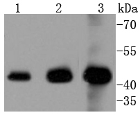
Western blot analysis of ERK1 on different lysates using anti-ERK1 antibody at 1/1,000 dilution. Positive control: Lane 1: Hela Lane 2: Jurkat Lane 3: K562
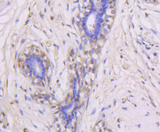
Immunohistochemical analysis of paraffin-embedded human breast tissue using anti-ERK1 antibody. Counter stained with hematoxylin.
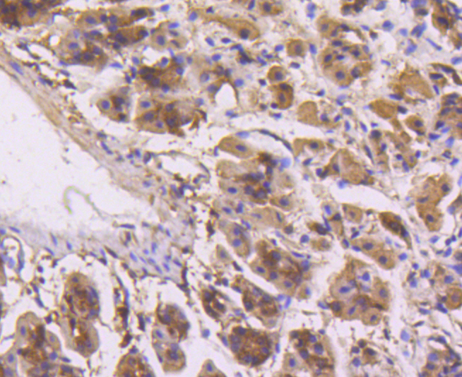
Immunohistochemical analysis of paraffin-embedded mouse stomach tissue using anti-ERK1 antibody. Counter stained with hematoxylin.
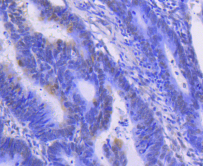
Immunohistochemical analysis of paraffin-embedded human colon cancer tissue using anti-ERK1 antibody. Counter stained with hematoxylin.
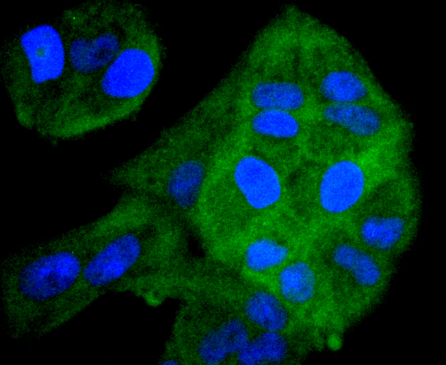
ICC staining ERK1 in Hela cells (green). The nuclear counter stain is DAPI (blue). Cells were fixed in paraformaldehyde, permeabilised with 0.25% Triton X100/PBS.
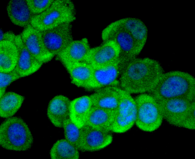
ICC staining ERK1 in MCF-7 cells (green). The nuclear counter stain is DAPI (blue). Cells were fixed in paraformaldehyde, permeabilised with 0.25% Triton X100/PBS.
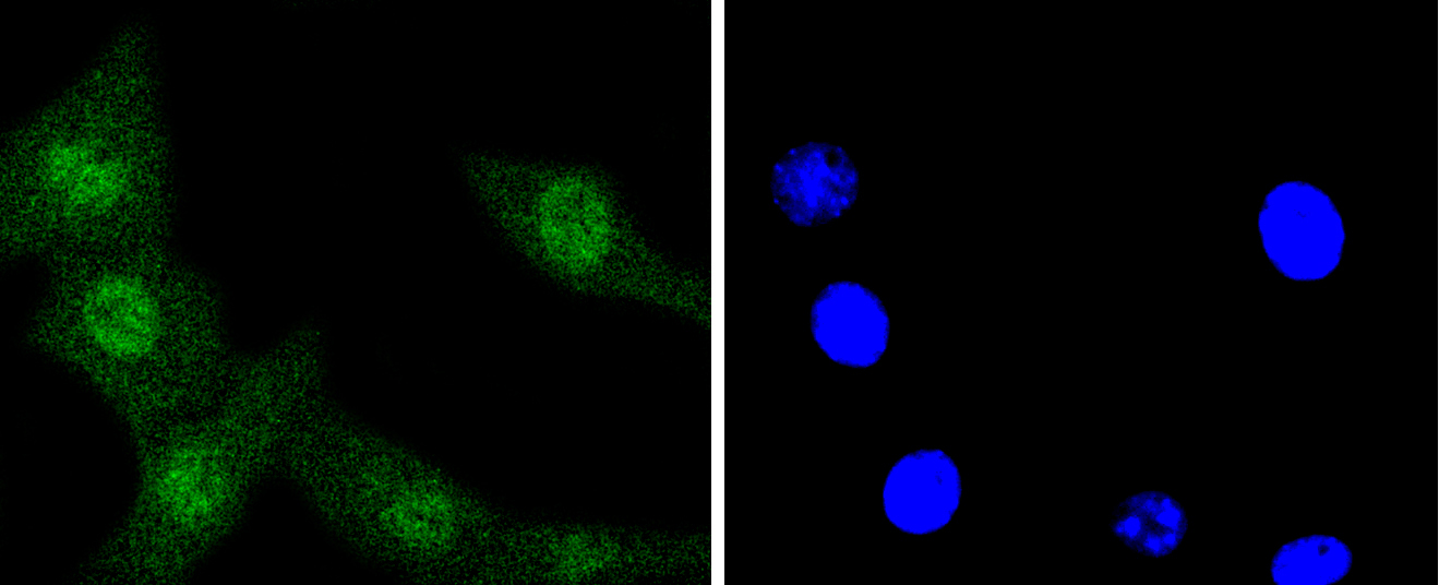
ICC staining ERK1 in NIH/3T3 cells (green). The nuclear counter stain is DAPI (blue). Cells were fixed in paraformaldehyde, permeabilised with 0.25% Triton X100/PBS.
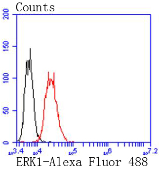
Flow cytometric analysis of Jurkat cells with ERK1 antibody at 1/50 dilution (red) compared with an unlabelled control (cells without incubation with primary antibody; black). Alexa Fluor 488-conjugated goat anti rabbit IgG was used as the secondary antibody.
Mitogen-activated protein kinase (MAPK) signaling pathways involve two closely related MAP kinases, known as extracellular-signal-related kinase 1 (ERK 1, p44) and 2 (ERK 2, p42). Growth factors, steroid hormones, G protein-coupled receptor ligands and neurotransmitters can initiate MAPK signaling pathways. Activation of ERK 1 and ERK 2 requires phosphorylation by upstream kinases such as MAP kinasekinase (MEK), MEK kinase and Raf-1. ERK 1 and ERK 2 phosphorylation can occur at specific tyrosine and threonine sites mapping within consensus motifs that include the threonine-glutamate-tyrosine motif. ERK activation leads to dimerization with other ERKs and subsequent localization to the nucleus. Active ERK dimers phosphorylate serine and threonine residues on nuclear proteins and influence a host of responses that include proliferation, differentiation, transcription regulation and development. The human ERK 1 gene maps to chromosome 16p12-p11.2 and encodes a 379 amino acid protein that shares 83% sequence identity to ERK 2.
If you have published an article using product 48709, please notify us so that we can cite your literature.


 Yes
Yes


