Product Detail
Product NamePHD1/prolyl hydroxylase Rabbit mAb
Clone No.SP00-48
Host SpeciesRecombinant Rabbit
Clonality Monoclonal
PurificationProA affinity purified
ApplicationsWB, ICC/IF, IHC, FC
Species ReactivityHu, Ms, Rt
Immunogen Descrecombinant protein
ConjugateUnconjugated
Other NamesDKFZp434E026 antibody
EGL nine (C.elegans) homolog 2 antibody
Egl nine homolog 2 (C. elegans) antibody
Egl nine homolog 2 antibody
EGLN 2 antibody
EGLN2 antibody
EGLN2_HUMAN antibody
EIT 6 antibody
EIT6 antibody
Estrogen-induced tag 6 antibody
HIF P4H 1 antibody
HIF PH1 antibody
HIF prolyl hydroxylase 1 antibody
HIF-PH1 antibody
HIF-prolyl hydroxylase 1 antibody
HIFPH 1 antibody
HIFPH1 antibody
HPH 3 antibody
HPH-1 antibody
HPH-3 antibody
HPH3 antibody
Hypoxia inducible factor prolyl hydroxylase 1 antibody
Hypoxia-inducible factor prolyl hydroxylase 1 antibody
P4H1 antibody
PHD 1 antibody
PhD1 antibody
prolyl hydroxylase domain containing protein 1 antibody
Prolyl hydroxylase domain-containing protein 1 antibody
Accession NoSwiss-Prot#:Q96KS0
Uniprot
Q96KS0
Gene ID
112398;
Calculated MW44 kDa
Formulation1*TBS (pH7.4), 1%BSA, 40%Glycerol. Preservative: 0.05% Sodium Azide.
StorageStore at -20˚C
Application Details
WB: 1:1,000-5,000
IHC: 1:50-1:200
ICC: 1:50-1:200
FC: 1:50-1:100
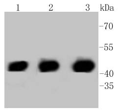
Western blot analysis of PHD1 on different lysates using anti-PHD1 antibody at 1/1,000 dilution. Positive control:
Lane 1: Hela
Lane 2: PC12
Lane 3: NIH/3T3
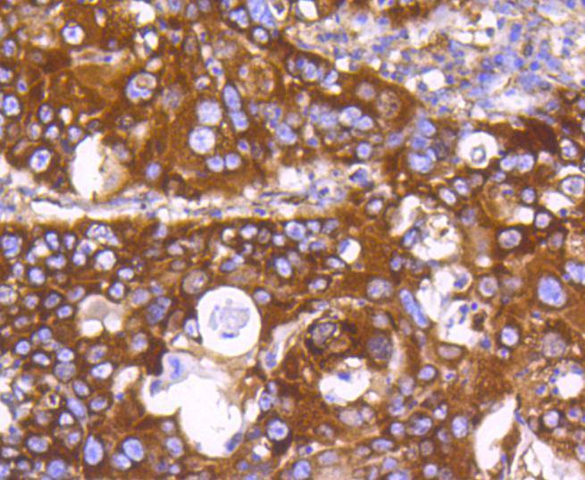
Immunohistochemical analysis of paraffin-embedded human lung cancer tissue using anti-PHD1 antibody. Counter stained with hematoxylin.
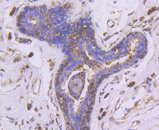
Immunohistochemical analysis of paraffin-embedded human breast carcinoma tissue using anti-PHD1 antibody. Counter stained with hematoxylin.
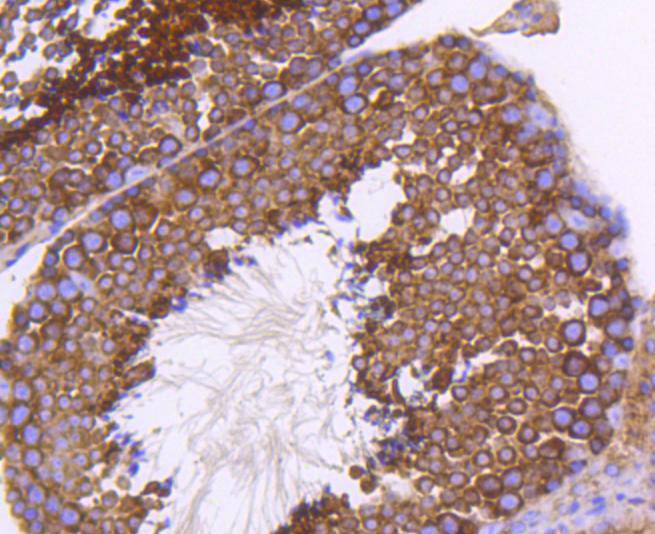
Immunohistochemical analysis of paraffin-embedded mouse testis tissue using anti-PHD1 antibody. Counter stained with hematoxylin.
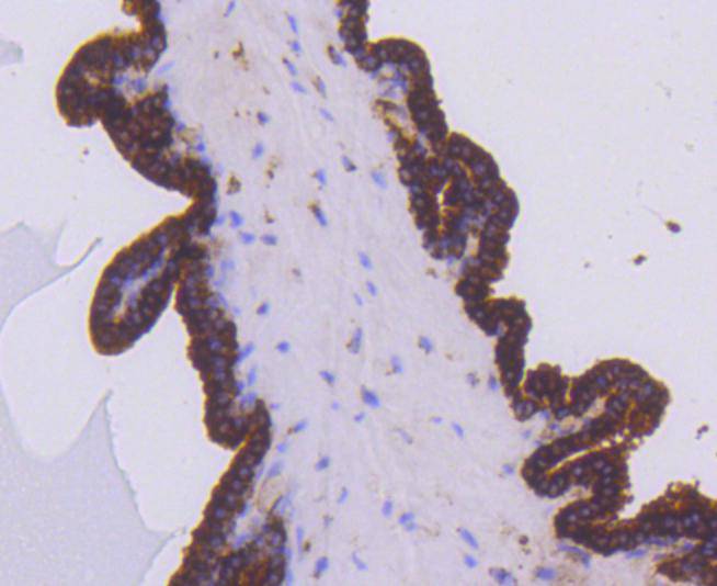
Immunohistochemical analysis of paraffin-embedded mouse prostate tissue using anti-PHD1 antibody. Counter stained with hematoxylin.
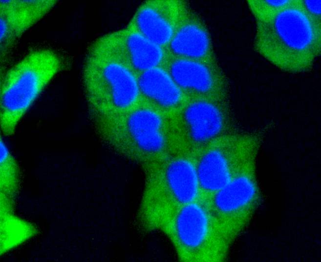
ICC staining PHD1 in Hela cells (green). The nuclear counter stain is DAPI (blue). Cells were fixed in paraformaldehyde, permeabilised with 0.25% Triton X100/PBS.
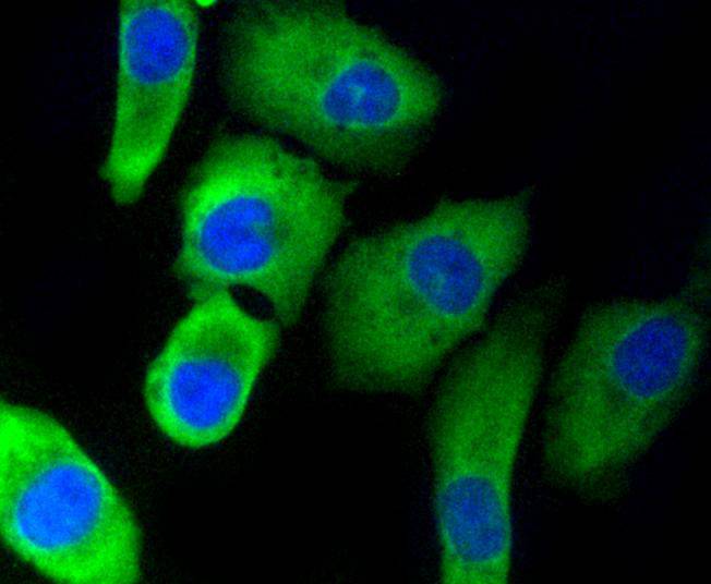
ICC staining PHD1 in A549 cells (green). The nuclear counter stain is DAPI (blue). Cells were fixed in paraformaldehyde, permeabilised with 0.25% Triton X100/PBS.
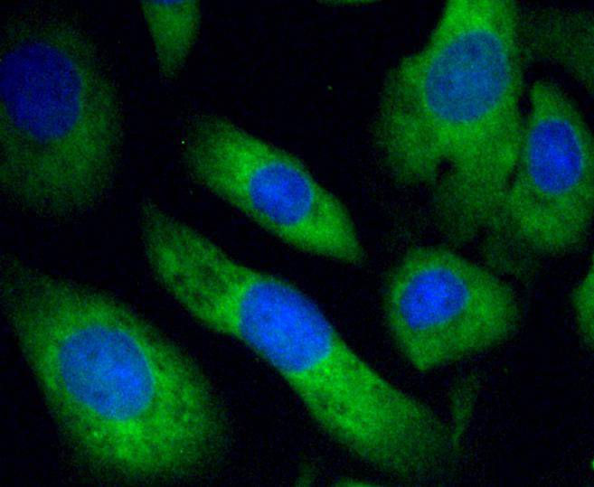
ICC staining PHD1 in SKOV-3 cells (green). The nuclear counter stain is DAPI (blue). Cells were fixed in paraformaldehyde, permeabilised with 0.25% Triton X100/PBS.
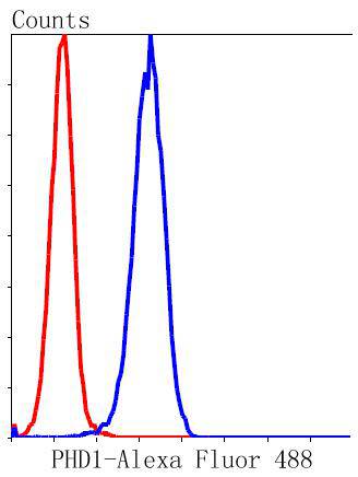
Flow cytometric analysis of Hela cells with PHD1 antibody at 1/50 dilution (blue) compared with an unlabelled control (cells without incubation with primary antibody; red). Alexa Fluor 488-conjugated goat anti rabbit IgG was used as the secondary antibody.
Prolyl hydroxylase domain proteins HIF PHD1, HIF PHD2 and HIF PHD3 (known as PHD1, PHD2 and PHD3 in rodents, respectively) can hydroxylate HIF-α subunits. Hypoxia-inducible factor (HIF) is a transcriptional regulator important in several aspects of oxygen homeostasis. The prolyl hydroxylases catalyze the posttranslational formation of 4-hydroxyproline in HIF-α proteins. HIF PHD1, which is widely expressed, with highest levels of expression in testis, functions as a cellular oxygen sensor and is important in cell growth regulation. HIF PHD1 can localize to the nucleus or the cytoplasm and is also detected in hormone responsive tissues, such as normal and cancerous mammary, ovarian and prostate epithelium. HIF PHD1 is encoded by EGLN2, which maps to chromosome 19q13.3. HIF PHD2 is regarded as the main cellular oxygen sensor, as RNA interference against HIF PHD2, but not HIF PHD1 or HIF PHD3, is enough to stabilize HIF-1α in normoxia. HIF PHD2, a direct HIF target gene, is expressed mainly in skeletal muscle, heart, kidney and brain. HIF PHD3 may play a role in the regulation of cell growth in muscle cells and in apoptosis in neuronal tissue. HIF PHD3 is widely expressed, although the highest levels can be detected in placenta and he
If you have published an article using product 48716, please notify us so that we can cite your literature.


 Yes
Yes


