Location:
Home
>
Recombinant Rabbit Monoclonal Antibodies > PMS2 Rabbit mAb
PMS2 Rabbit mAb#48717

 Yes
Yes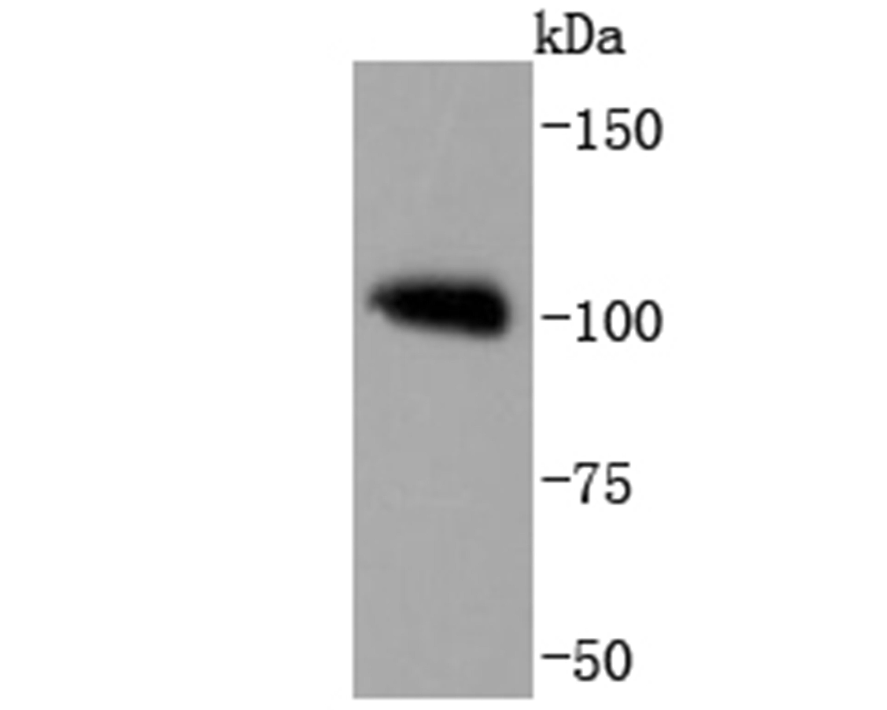
Western blot analysis of PMS2 on Hela cell lysates. Proteins were transferred to a PVDF membrane and blocked with 5% BSA in PBS for 1 hour at room temperature. The primary antibody was used at a 1:1,000 dilution in 5% BSA at room temperature for 2 hours. Goat Anti-Mouse IgG - HRP Secondary Antibody (HA1006) at 1:5,000 dilution was used for 1 hour at room temperature.
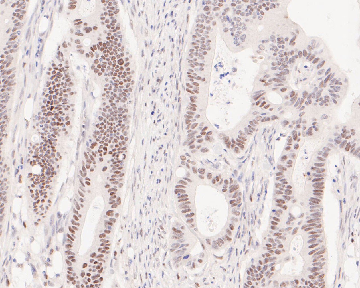
Immunohistochemical analysis of paraffin-embedded human colon cancer tissue using anti-PMS2 antibody. The section was pre-treated using heat mediated antigen retrieval with sodium citrate buffer (pH6) for 20 mins. The tissues were blocked in 5% BSA for 30 minutes at room temperature, washed with ddH2O and PBS, and then probed with the antibody (ET1605-1) at 1/200 dilution, for 30 minutes at room temperature and detected using an HRP conjugated compact polymer system. DAB was used as the chrogen. Counter stained with hematoxylin and mounted with DPX.
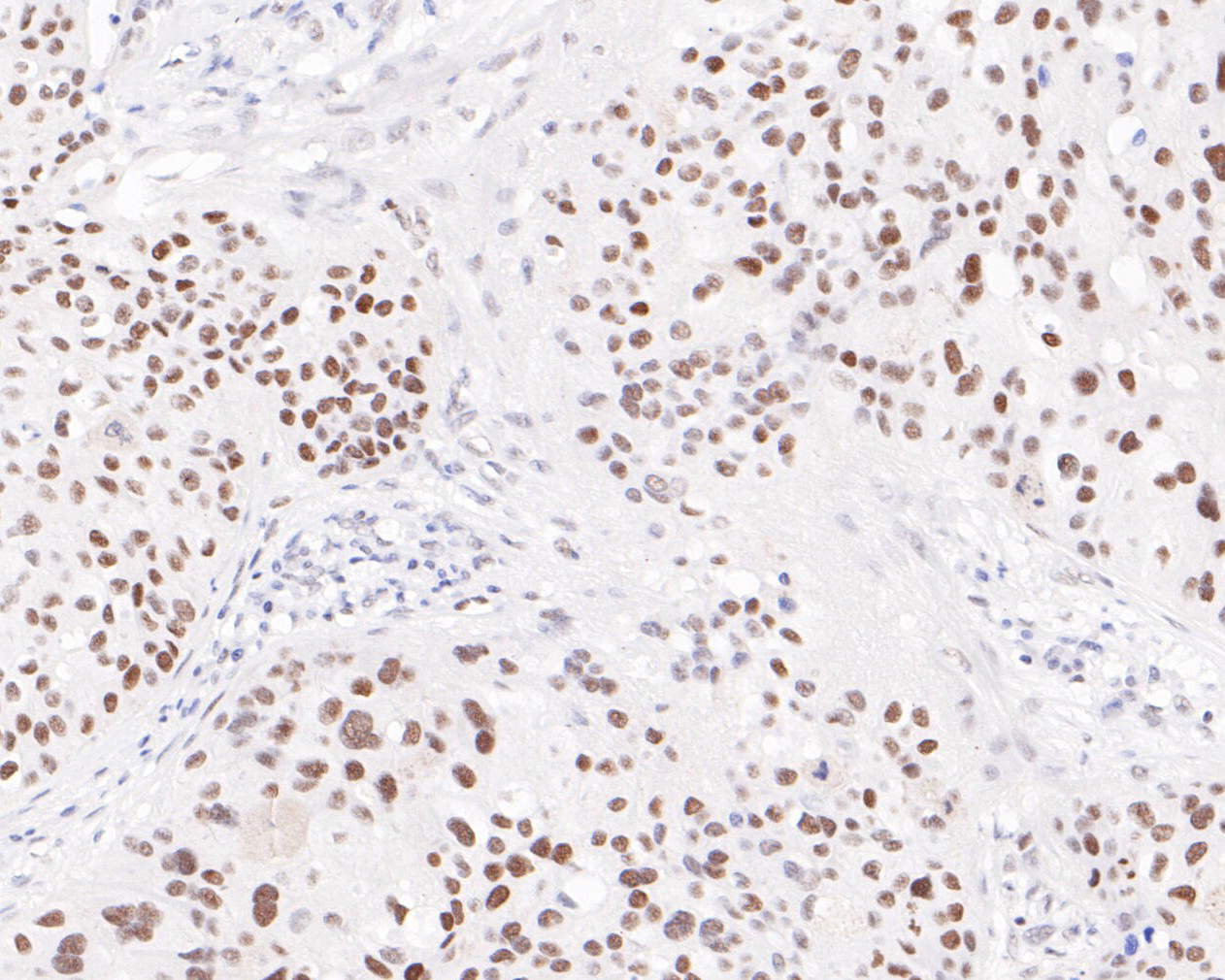
Immunohistochemical analysis of paraffin-embedded human breast cancer tissue using anti-PMS2 antibody. The section was pre-treated using heat mediated antigen retrieval with sodium citrate buffer (pH6) for 20 mins. The tissues were blocked in 5% BSA for 30 minutes at room temperature, washed with ddH2O and PBS, and then probed with the antibody (ET1605-1) at 1/200 dilution, for 30 minutes at room temperature and detected using an HRP conjugated compact polymer system. DAB was used as the chrogen. Counter stained with hematoxylin and mounted with DPX.
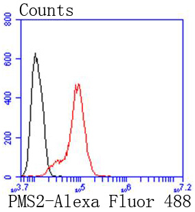
Flow cytometric analysis of PMS2 was done on Hela cells. The cells were fixed, permeabilized and stained with APE1 antibody at 1/100 dilution (red) compared with an unlabelled control (cells without incubation with primary antibody; black). After incubation of the primary antibody on room temperature for an hour, the cells was stained with a Alexa Fluor? 488-conjugated goat anti-rabbit IgG Secondary antibody at 1/500 dilution for 30 minutes.

ICC staining PMS2 in Hela cells (red). Formalin fixed cells were permeabilized with 0.1% Triton X-100 in TBS for 10 minutes at room temperature and blocked with 1% Blocker BSA for 15 minutes at room temperature. Cells were probed with Carbonic anhydrase 2 monoclonal antibody at a dilution of 1:100 for at least 1 hour at room temperature, washed with PBS. Alexa Fluorc? 488 Goat anti-Mouse IgG was used as the secondary antibody at 1/100 dilution. The nuclear counter stain is DAPI (blue).
NOTE
Application
- WBWestern Blotting
- IHCImmunohistochemistry
- IFImmunofluorescence
- ICCImmunocytochemistry
- FCFlow Cytometry
- IPImmunoprecipitation
- EELISA
- DBDot Blotting
- ChIPChromatin Immunoprecipitation
- GICAGold Immunochromatography Assay
- NCNegative Control
Species Reactivity
- HuHuman
- MsMouse
- RtRat
- DmDrosophila melanogaster
- CCaenorhabditis elegans
- MkMonkey
- RbRabbit
- BBovine
- DDog
- PPig
- HmHamster
- ChHmChinese Hamster
- ChkChicken
- ShpSheep


