Welcome to Sabbiotech!     
Email:info@signalwayantibody.com
Location: Home > Recombinant Rabbit Monoclonal Antibodies > STAT3 Rabbit mAb

STAT3 Rabbit mAb#48732

Availability: Yes
Total Amount:$
Product Detail

Product NameSTAT3 Rabbit mAb

Clone No.SY34-01

Host SpeciesRecombinant Rabbit

Clonality Monoclonal

PurificationProA affinity purified

ApplicationsWB, ICC/IF, IHC

Species ReactivityHu, Ms, Rt

Immunogen Descrecombinant protein

ConjugateUnconjugated

Other Names1110034C02Rik antibody
Acute Phase Response Factor antibody
Acute-phase response factor antibody
ADMIO antibody
APRF antibody
AW109958 antibody
DNA binding protein APRF antibody
FLJ20882 antibody
HIES antibody
MGC16063 antibody
Signal transducer and activator of transcription 3 (acute phase response factor) antibody
Signal transducer and activator of transcription 3 antibody
STAT 3 antibody
Stat3 antibody
STAT3_HUMAN antibody

Accession NoSwiss-Prot#:P40763

Uniprot P40763

Gene ID 6774;

Calculated MW88 kDa

Formulation1*TBS (pH7.4), 1%BSA, 40%Glycerol. Preservative: 0.05% Sodium Azide.

StorageStore at -20˚C

Application Details
WB: 1:1,000-1:2,000
IHC: 1:50-1:200
ICC: 1:100-1:500

Western blot analysis of STAT3 on MCF-7 cells lysates using anti-STAT3 antibody at 1/1,000 dilution.
Immunohistochemical analysis of paraffin-embedded human gastric carcinoma tissue using anti-STAT3 antibody. Counter stained with hematoxylin.
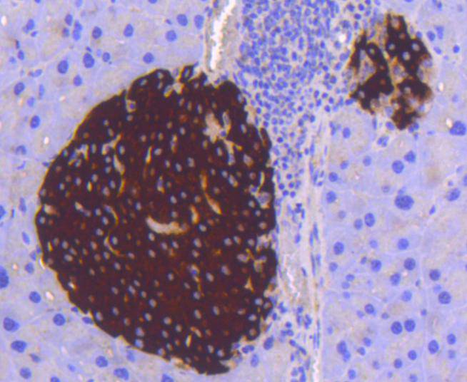
Immunohistochemical analysis of paraffin-embedded mouse pancreas tissue using anti-STAT3 antibody. Counter stained with hematoxylin.
Immunohistochemical analysis of paraffin-embedded human breast carcinoma tissue using anti-STAT3 antibody. Counter stained with hematoxylin.
Immunohistochemical analysis of paraffin-embedded mouse brain tissue using anti-STAT3 antibody. Counter stained with hematoxylin.
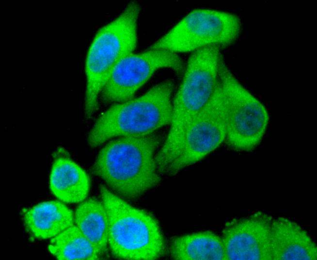
ICC staining STAT3 in Hela cells (green). The nuclear counter stain is DAPI (blue). Cells were fixed in paraformaldehyde, permeabilised with 0.25% Triton X100/PBS.
ICC staining STAT3 in A549 cells (green). The nuclear counter stain is DAPI (blue). Cells were fixed in paraformaldehyde, permeabilised with 0.25% Triton X100/PBS.
ICC staining STAT3 in HepG2 cells (green). The nuclear counter stain is DAPI (blue). Cells were fixed in paraformaldehyde, permeabilised with 0.25% Triton X100/PBS.
Membrane receptor signaling by various ligands, including interferons and growth hormones such as EGF, induces activation of JAK kinases which then leads to tyrosine phosphorylation of the various Stat transcription factors. Stat1 and Stat2 are induced by IFN-α and form a heterodimer which is part of the ISGF3 transcription factor complex. Although early reports indicate Stat3 activation by EGF and IL-6, it has been shown that Stat3β appears to be activated by both while Stat3α is activated by EGF, but not by IL-6. Highest expression of Stat4 is seen in testis and myeloid cells. IL-12 has been identified as an activator of Stat4. Stat5 has been shown to be activated by Prolactin and by IL-3. Stat6 is involved in IL-4 activated signaling pathways.

If you have published an article using product 48732, please notify us so that we can cite your literature.

NOTE

Application

  • WBWestern Blotting
  • IHCImmunohistochemistry
  • IFImmunofluorescence
  • ICCImmunocytochemistry
  • FCFlow Cytometry
  • IPImmunoprecipitation
  • EELISA
  • DBDot Blotting
  • ChIPChromatin Immunoprecipitation
  • GICAGold Immunochromatography Assay
  • NCNegative Control

Species Reactivity

  • HuHuman
  • MsMouse
  • RtRat
  • DmDrosophila melanogaster
  • CCaenorhabditis elegans
  • MkMonkey
  • RbRabbit
  • BBovine
  • DDog
  • PPig
  • HmHamster
  • ChHmChinese Hamster
  • ChkChicken
  • ShpSheep
;