Product Detail
Product NameNGF Rabbit mAb
Clone No.SI79-01
Host SpeciesRecombinant Rabbit
Clonality Monoclonal
PurificationProA affinity purified
ApplicationsWB, ICC/IF, IHC
Species ReactivityHu, Ms, Rt, zebrafish
Immunogen Descrecombinant protein
ConjugateUnconjugated
Other NamesBeta nerve growth factor antibody Beta NGF antibody Beta-nerve growth factor antibody Beta-NGF antibody HSAN5 antibody MGC161426 antibody MGC161428 antibody Nerve growth factor (beta polypeptide) antibody Nerve growth factor antibody Nerve growth factor beta antibody Nerve growth factor beta polypeptide antibody Nerve growth factor beta subunit antibody NGF antibody NGF_HUMAN antibody NGFB antibody NID67 antibody
Accession NoSwiss-Prot#:P01138
Uniprot
P01138
Gene ID
4803;
Calculated MW32 kDa
Formulation1*TBS (pH7.4), 1%BSA, 40%Glycerol. Preservative: 0.05% Sodium Azide.
StorageStore at -20˚C
Application Details
WB: 1:1,000-1:2,000
IHC: 1:50-1:200
ICC: 1:50-1:200
Western blot analysis of NGF on Hela cell lysates using anti-NGF antibody at 1/1,000 dilution.
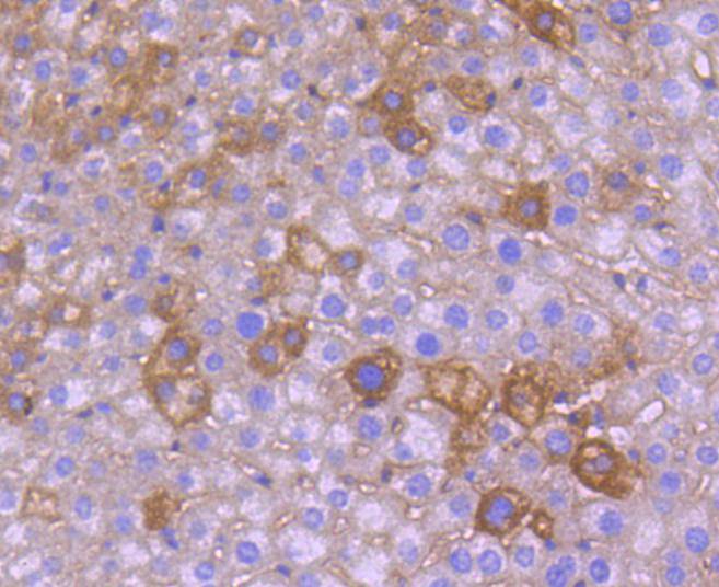
Immunohistochemical analysis of paraffin-embedded mouse liver tissue using anti-NGF antibody. Counter stained with hematoxylin.
Immunohistochemical analysis of paraffin-embedded mouse brain tissue using anti-NGF antibody. Counter stained with hematoxylin.
Immunohistochemical analysis of paraffin-embedded mouse thymus tissue using anti-NGF antibody. Counter stained with hematoxylin.
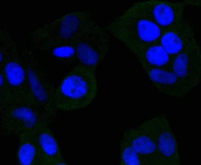
ICC staining NGF in Hela cells (green). The nuclear counter stain is DAPI (blue). Cells were fixed in paraformaldehyde, permeabilised with 0.25% Triton X100/PBS.
ICC staining NGF in HepG2 cells (green). The nuclear counter stain is DAPI (blue). Cells were fixed in paraformaldehyde, permeabilised with 0.25% Triton X100/PBS.
ICC staining NGF in NIH/3T3 cells (green). The nuclear counter stain is DAPI (blue). Cells were fixed in paraformaldehyde, permeabilised with 0.25% Triton X100/PBS.
Neurotrophins function to regulate naturally occurring cell death of neurons during development. The prototype neurotrophin is nerve growth factor (NGF), originally discovered in the 1950s as a soluble peptide promoting the survival of, and neurite outgrowth from, sympathetic ganglia. Three additional structurally homologous neurotrophic factors have been identified. These include brain-derived neurotrophic factor (BDNF), neurotrophin-3 (NT-3) and neurotrophin-4 (NT-4) (also designated NT-5). These various neurotrophins stimulate the in vitro survival of distinct, but partially overlapping, populations of neurons. The cell surface receptors through which neurotrophins mediate their activity have been identified. For instance, the Trk A receptor is the preferential receptor for NGF, but also binds NT-3 and NT-4. The Trk B receptor binds both BDNF and NT-4 equally well, and binds NT-3 to a lesser extent, while the Trk C receptor only binds NT-3.
If you have published an article using product 48748, please notify us so that we can cite your literature.


 Yes
Yes


