Product Detail
Product NameMet(C-Met) Rabbit mAb
Clone No.SJ19-05
Host SpeciesRecombinant Rabbit
Clonality Monoclonal
PurificationProA affinity purified
ApplicationsWB, ICC/IF, IHC, FC
Species ReactivityHu
Immunogen Descrecombinant protein
ConjugateUnconjugated
Other NamesAUTS9 antibody c met antibody D249 antibody Hepatocyte growth factor receptor antibody HGF antibody HGF receptor antibody HGF/SF receptor antibody HGFR antibody MET antibody Met proto oncogene tyrosine kinase antibody MET proto oncogene, receptor tyrosine kinase antibody Met proto-oncogene (hepatocyte growth factor receptor) antibody Met proto-oncogene antibody Met protooncogene antibody MET_HUMAN antibody Oncogene MET antibody Par4 antibody Proto-oncogene c-Met antibody RCCP2 antibody Scatter factor receptor antibody SF receptor antibody Tyrosine-protein kinase Met antibody
Accession NoSwiss-Prot#:P08581
Uniprot
P08581
Gene ID
4233;
Calculated MW155 kDa
Formulation1*TBS (pH7.4), 1%BSA, 40%Glycerol. Preservative: 0.05% Sodium Azide.
StorageStore at -20˚C
Application Details
WB: 1:1,000-5,000
IHC: 1:50-1:200
ICC: 1:50-1:200
FC: 1:50-1:100
Western blot analysis of Met on different lysates using anti-Met antibody at 1/1,000 dilution. Positive control: Lane 1: Hela Lane 2: HepG2
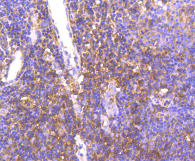
Immunohistochemical analysis of paraffin-embedded human tonsil tissue using anti-Met antibody. Counter stained with hematoxylin.
Immunohistochemical analysis of paraffin-embedded human lung cancer tissue using anti-Met antibody. Counter stained with hematoxylin.
Immunohistochemical analysis of paraffin-embedded human liver cancer tissue using anti-Met antibody. Counter stained with hematoxylin.
Immunohistochemical analysis of paraffin-embedded human breast carcinoma tissue using anti-Met antibody. Counter stained with hematoxylin.
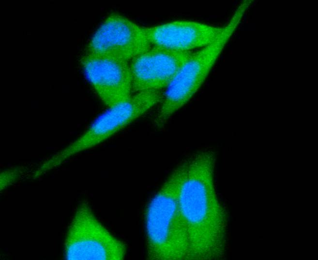
ICC staining Met in Hela cells (green). The nuclear counter stain is DAPI (blue). Cells were fixed in paraformaldehyde, permeabilised with 0.25% Triton X100/PBS.
Flow cytometric analysis of Hela cells with Met antibody at 1/50 dilution (red) compared with an unlabelled control (cells without incubation with primary antibody; black). Alexa Fluor 488-conjugated goat anti rabbit IgG was used as the secondary antibody.
The c-Met oncogene was originally isolated from a chemical carcinogen-treated human osteogenic sarcoma cell line by transfection analysis in NIH/3T3 cells. The Met proto-oncogene product was identified as a transmembrane receptor-like protein with tyrosine kinase activity that is expressed in many tissues. A high proportion of spontaneous NIH/3T3 transformants overexpress c-Met and by transfection analysis the c-Met proto-oncogene has been shown to exhibit transforming activity. Tyrosine phosphorylation of apparently normal Met protein has also been observed in certain human gastric carcinoma cell lines . The c-Met gene product has been identified as the cell-surface receptor for hepatocyte growth factor, a plasminogen-like protein thought to be a humoral mediator of liver regeneration.
If you have published an article using product 48758, please notify us so that we can cite your literature.


 Yes
Yes


