Welcome to Sabbiotech!     
Email:info@signalwayantibody.com
Location: Home > Recombinant Rabbit Monoclonal Antibodies > Calmodulin Rabbit mAb

Calmodulin Rabbit mAb#48759

Availability: Yes
Total Amount:$
Product Detail

Product NameCalmodulin Rabbit mAb

Clone No.SJ16-09

Host SpeciesRecombinant Rabbit

Clonality Monoclonal

PurificationProA affinity purified

ApplicationsWB, ICC/IF, IHC, IP

Species ReactivityHu, Ms, Rt

Immunogen Descrecombinant protein

ConjugateUnconjugated

Other NamesCALM 1 antibody
CALM 2 antibody
CALM 3 antibody
CALM antibody
CALM_HUMAN antibody
CALM1 antibody
CALM2 antibody
Calm3 antibody
CALML2 antibody
calmodulin 1 (phosphorylase kinase, delta) antibody
Calmodulin 2 (phosphorylase kinase, delta) antibody
Calmodulin 3 (phosphorylase kinase, delta) antibody
Calmodulin antibody
CaM antibody
CAM I antibody
CAM1 antibody
CAM2 antibody
CAM3 antibody
CAMB antibody
CAMC antibody
CAMI antibody
CAMII antibody
CPVT4 antibody
DD132 antibody
FLJ99410 antibody
LP7057 protein antibody
PHKD antibody
PHKD2 antibody
PHKD3 antibody
phosphorylase kinase delta antibody
phosphorylase kinase, delta subunit antibody

Accession NoSwiss-Prot:P62158
Gene ID:801

Uniprot P62158

Gene ID 801;

Calculated MW17 kDa

Formulation1*TBS (pH7.4), 1%BSA, 40%Glycerol. Preservative: 0.05% Sodium Azide.

StorageStore at -20˚C

Application Details
WB: 1:1,000
IHC: 1:50-1:200
ICC: 1:50-1:200

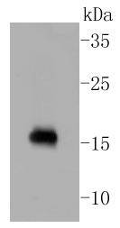
Western blot analysis of Calmodulin on rat brain lysates using anti-Calmodulin antibody at 1/1,000 dilution.
Immunohistochemical analysis of paraffin-embedded mouse brain tissue using anti-Calmodulin antibody. Counter stained with hematoxylin.
Immunohistochemical analysis of paraffin-embedded mouse cerebellum tissue using anti-Calmodulin antibody. Counter stained with hematoxylin.
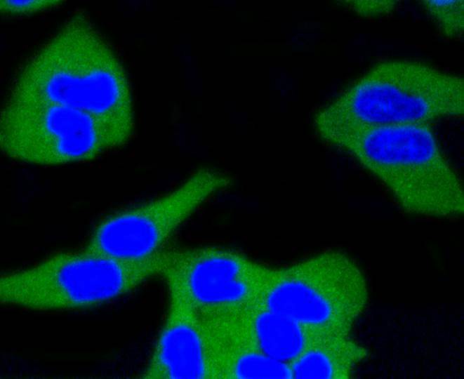
ICC staining Calmodulin in Hela cells (green). The nuclear counter stain is DAPI (blue). Cells were fixed in paraformaldehyde, permeabilised with 0.25% Triton X100/PBS.
ICC staining Calmodulin in MCF-7 cells (green). The nuclear counter stain is DAPI (blue). Cells were fixed in paraformaldehyde, permeabilised with 0.25% Triton X100/PBS.
ICC staining Calmodulin in NIH/3T3 cells (green). Cells were fixed in paraformaldehyde, permeabilised with 0.25% Triton X100/PBS.
The level of intracellular calcium is tightly regulated in all eukaryotic cells. A modest increase in this level can result in a myriad of physiological responses, most of which are mediated by calmodulin (CaM), the universal calcium sensor. CaM directly modulates the activity of protein kinases and phosphatases, ion channels and nitric oxide synthetases. It is generally involved in such diverse processes as cell proliferation, endocytosis, cellular adhesion, protein turn over and smooth muscle contraction. CaM (calmodulin) is an acidic protein, 148 amino acids in length, with four helix-loop-helix calcium binding domains. In humans, 3 distinct genes have been identified (CALM1, CALM2 and CALM3); each encoding the identical protein. CALML3 (calmodulin-like 3, or calmodulin-related protein NB-1) shares significant sequence identity with CaM and it is suggested that it may competitively bind CaM substrates. Interestingly, CaM has been shown to associate with the carboxy terminus of the dystrophin gene product, implying that it may regulate its activity.

If you have published an article using product 48759, please notify us so that we can cite your literature.

NOTE

Application

  • WBWestern Blotting
  • IHCImmunohistochemistry
  • IFImmunofluorescence
  • ICCImmunocytochemistry
  • FCFlow Cytometry
  • IPImmunoprecipitation
  • EELISA
  • DBDot Blotting
  • ChIPChromatin Immunoprecipitation
  • GICAGold Immunochromatography Assay
  • NCNegative Control

Species Reactivity

  • HuHuman
  • MsMouse
  • RtRat
  • DmDrosophila melanogaster
  • CCaenorhabditis elegans
  • MkMonkey
  • RbRabbit
  • BBovine
  • DDog
  • PPig
  • HmHamster
  • ChHmChinese Hamster
  • ChkChicken
  • ShpSheep
;