Welcome to Sabbiotech!     
Email:info@signalwayantibody.com
Location: Home > Recombinant Rabbit Monoclonal Antibodies > CD3 epsilon Rabbit mAb

CD3 epsilon Rabbit mAb#48775

Availability: Yes
Total Amount:$
Product Detail

Product NameCD3 epsilon Rabbit mAb

Clone No.SR4520

Host SpeciesRecombinant Rabbit

ClonalityMonoclonal

PurificationProA affinity purified

ApplicationsWB, IHC, IP, FC

Species ReactivityHuman Mouse Rat

Immunogen Descrecombinant protein

ConjugateUnconjugated

Other NamesCD3 epsilon antibody
CD3e antibody
CD3e antigen epsilon polypeptide (TiT3 complex) antibody
CD3E antigen epsilon polypeptide antibody
CD3E antigen, epsilon subunit antibody
CD3e molecule epsilon antibody
CD3e molecule, epsilon (CD3 TCR complex) antibody
CD3e molecule, epsilon (CD3-TCR complex) antibody
CD3E_HUMAN antibody
IMD18 antibody
T cell antigen receptor complex epsilon subunit of T3 antibody
T cell surface antigen T3/Leu 4 epsilon chain antibody
T cell surface glycoprotein CD3 epsilon chain antibody
T-cell surface antigen T3/Leu-4 epsilon chain antibody
T-cell surface glycoprotein CD3 epsilon chain antibody
T3E antibody
TCRE antibody

Accession NoSwiss-Prot#:P07766

Uniprot P07766

Gene ID 916;

Calculated MW23 kDa

Formulation1*TBS (pH7.4), 1%BSA, 40%Glycerol. Preservative: 0.05% Sodium Azide.

StorageStore at -20˚C

Application Details
WB: 1:1,000-1:2,000
IHC:1:50-1:200


FC: 1:50-1:100
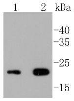
Western blot analysis of CD3 epsilon on different lysates using anti-CD3 epsilon antibody at 1/1,000 dilution. Positive control:
Lane 1: Jurkat
Lane 2: Human thymus
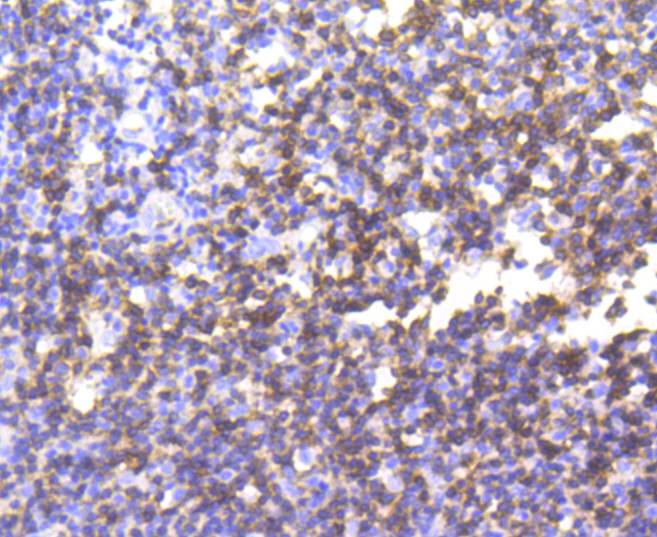
Immunohistochemical analysis of paraffin-embedded human tonsil tissue using anti-CD3 epsilon antibody. Counter stained with hematoxylin.
Immunohistochemical analysis of paraffin-embedded human spleen tissue using anti-CD3 epsilon antibody. Counter stained with hematoxylin.
Flow cytometric analysis of Jurkat cells with CD3 epsilon antibody at 1/50 dilution (blue) compared with an unlabelled control (cells without incubation with primary antibody; red). Alexa Fluor 488-conjugated goat anti rabbit IgG was used as the secondary antibody
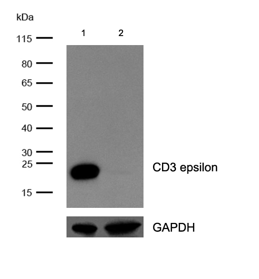
All lanes : CD3 epsilon Rabbit mAb at 1/2k dilution
Lane 1 : Wild-type Jurkat cell lysate
Lane 2 : CD3 epsilon knockout Jurkat cell lysate
Lysates/proteins at 20 µg per lane.
The T cell antigen receptor (TCR) recognizes foreign antigens and translates such recognition events into intracellular signals that elicit a change in the cell from a dormant to an activated state. Much of this signaling process can be attributed to a multisubunit complex of proteins that associates directly with the TCR. This complex has been designated CD3 (cluster of differentiation 3). CD3 is composed of four polypeptides: ζ, γ, ε and δ. Each of these polypeptides contains at least one immunoreceptor tyrosine-based activation motif (ITAM). Engagement of TCR complex with foreign antigens induces tyrosine phosphorylation in the ITAM motifs and phosphorylated ITAMs function as docking sites for signaling molecules such as ZAP-70 and p85 subunit of PI-3 kinase. TCR ligation also induces a conformational change in CD3ε, such that a proline region is exposed and then associates with the adaptor protein Nck.

If you have published an article using product 48775, please notify us so that we can cite your literature.

NOTE

Application

  • WBWestern Blotting
  • IHCImmunohistochemistry
  • IFImmunofluorescence
  • ICCImmunocytochemistry
  • FCFlow Cytometry
  • IPImmunoprecipitation
  • EELISA
  • DBDot Blotting
  • ChIPChromatin Immunoprecipitation
  • GICAGold Immunochromatography Assay
  • NCNegative Control

Species Reactivity

  • HuHuman
  • MsMouse
  • RtRat
  • DmDrosophila melanogaster
  • CCaenorhabditis elegans
  • MkMonkey
  • RbRabbit
  • BBovine
  • DDog
  • PPig
  • HmHamster
  • ChHmChinese Hamster
  • ChkChicken
  • ShpSheep
;