Product Detail
Product NameCytokeratin 8+18 Rabbit mAb
Clone No.SU0338
Host SpeciesRecombinant Rabbit
Clonality Monoclonal
PurificationProA affinity purified
ApplicationsWB, ICC/IF, IHC, IP, FC
Species ReactivityHu, Ms
Immunogen Descrecombinant protein
ConjugateUnconjugated
Other NamesCARD2 antibody Cell proliferation inducing gene 46 protein antibody Cell proliferation inducing protein 46 antibody CK 8 antibody CK-8 antibody CK18 antibody CK8 antibody CYK18 antibody CYK8 antibody Cytokeratin 18 antibody Cytokeratin 8 antibody Cytokeratin-8 antibody K18 antibody K2C8 antibody K2C8_HUMAN antibody K8 antibody Keratin 18 antibody Keratin 8 antibody Keratin antibody Keratin type I cytoskeletal 18 antibody keratin type II cytoskeletal 8 antibody Keratin-8 antibody KO antibody KRT18 antibody KRT8 antibody type II cytoskeletal 8 antibody Type-II keratin Kb8 antibody
Accession NoSwiss-Prot#:P05783
Uniprot
P05783
Gene ID
3875;
Calculated MW53 kDa
Formulation1*TBS (pH7.4), 1%BSA, 40%Glycerol. Preservative: 0.05% Sodium Azide.
StorageStore at -20˚C
Application Details
WB: 1:1,000-1:2,000
IHC: 1:50-1:200
ICC: 1:50-1:200
FC: 1:50-1:100
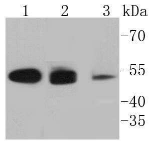
Western blot analysis of Cytokeratin 8+18 on different lysates using anti-Cytokeratin 8+18 antibody at 1/1,000 dilution. Positive control: Lane 1: Hela Lane 2: MCF-7 Lane 3: A431
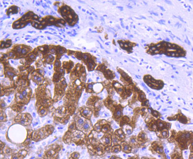
Immunohistochemical analysis of paraffin-embedded human liver tissue using anti-Cytokeratin 8+18 antibody. Counter stained with hematoxylin.
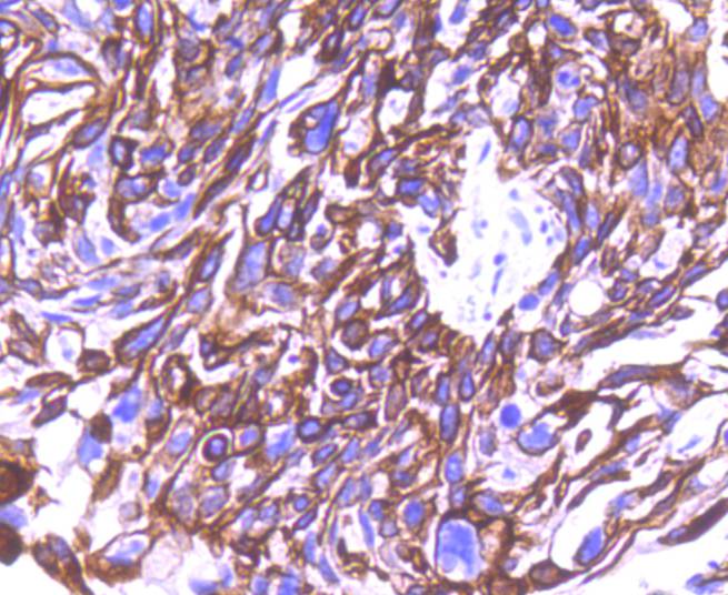
Immunohistochemical analysis of paraffin-embedded human breast carcinoma tissue using anti-Cytokeratin 8+18 antibody. Counter stained with hematoxylin.
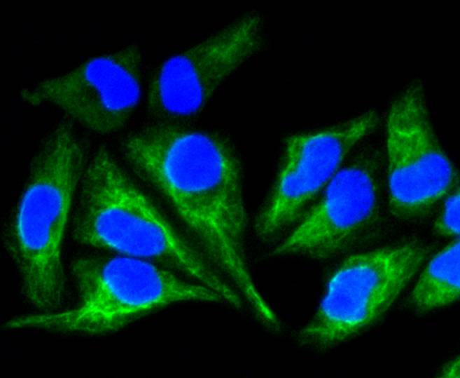
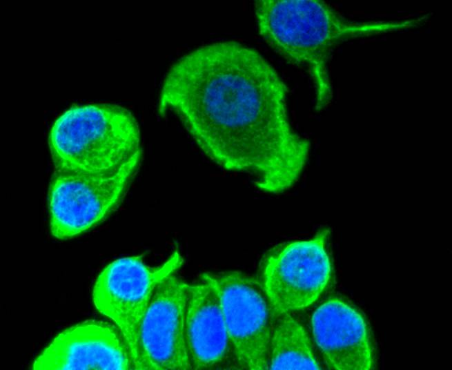
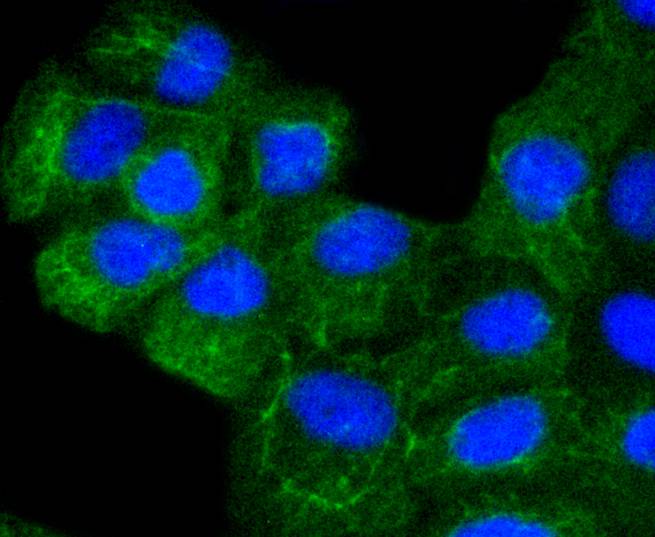
ICC staining Cytokeratin 8+18 in Hela cells (green). The nuclear counter stain is DAPI (blue). Cells were fixed in paraformaldehyde, permeabilised with 0.25% Triton X100/PBS.
ICC staining Cytokeratin 8+18 in MCF-7 cells (green). The nuclear counter stain is DAPI (blue). Cells were fixed in paraformaldehyde, permeabilised with 0.25% Triton X100/PBS.
ICC staining Cytokeratin 8+18 in A431 cells (green). The nuclear counter stain is DAPI (blue). Cells were fixed in paraformaldehyde, permeabilised with 0.25% Triton X100/PBS.
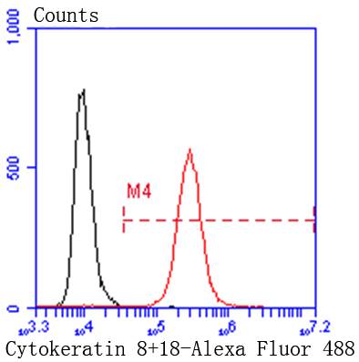
Flow cytometric analysis of Hela cells with Cytokeratin 8+18 antibody at 1/50 dilution (red) compared with an unlabelled control (cells without incubation with primary antibody; black). Alexa Fluor 488-conjugated goat anti rabbit IgG was used as the secondary antibody.
Cytokeratins comprise a diverse group of intermediate filament proteins (IFPs) that are expressed as pairs in both keratinized and non-keratinized epithelial tissue. Cytokeratins play a critical role in differentiation and tissue specialization and function to maintain the overall structural integrity of epithelial cells. They have been found to be useful markers of tissue differentiation, which is directly applicable to the characterization of malignant tumors. Cytokeratin 8 expression is seen in epithelium and epithelium-derived tumors. The Cytokeratin 8 and 18 pair are normally expressed in simple epithelia, but not in stratified epithelial cells. Research indicates that squamous cell carcinomas derived from stratified epithelia show abnormal expression of Cytokeratin 8 and 18, although it is not known whether these proteins contribute to the malignant phenotype of the cells. Expression of Cytokeratin 8 and 18 in oral squamous cell carcinomas is an independent prognostic marker that indicates a poor prognosis. Cytokeratin 8 expression correlates with malignancy in leukoplakia and carcinomas of the head and neck; it is expressed in all non-small-cell lung cancers. Cytokeratin 8 has been shown to possess extracellular epitopes on tumor cells, which may represent valuable targets for therapy.
If you have published an article using product 48822, please notify us so that we can cite your literature.


 Yes
Yes


