Product Detail
Product Namep27 KIP 1 Rabbit mAb
Clone No.SU37-04
Host SpeciesRecombinant Rabbit
Clonality Monoclonal
PurificationProA affinity purified
ApplicationsWB, ICC/IF, IHC, IP, FC
Species ReactivityHu, Rt
Immunogen Descrecombinant protein
ConjugateUnconjugated
Other NamesAA408329 antibody AI843786 antibody Cdki1b antibody CDKN 1B antibody CDKN 4 antibody CDKN1B antibody CDKN4 antibody CDN1B_HUMAN antibody Cyclin Dependent Kinase Inhibitor 1B antibody Cyclin dependent kinase inhibitor p27 antibody Cyclin-dependent kinase inhibitor 1B (p27, Kip1) antibody Cyclin-dependent kinase inhibitor 1B antibody Cyclin-dependent kinase inhibitor p27 antibody Cyclin-dependent kinase inhibitor p27 Kip1 antibody KIP 1 antibody KIP1 antibody MEN1B antibody MEN4 antibody OTTHUMP00000195098 antibody OTTHUMP00000195099 antibody p27 antibody p27 Kip1 antibody P27-like cyclin-dependent kinase inhibitor antibody p27Kip1 antibody
Accession NoSwiss-Prot#:P46527
Uniprot
P46527
Gene ID
1027;
Calculated MW27 kDa
Formulation1*TBS (pH7.4), 1%BSA, 40%Glycerol. Preservative: 0.05% Sodium Azide.
StorageStore at -20˚C
Application Details
WB: 1:1,000
IHC: 1:100-1:500
ICC: 1:100-1:500
FC: 1:50-1:100
Immunohistochemical analysis of paraffin-embedded human tonsil tissue using anti-p27 KIP 1 antibody. Counter stained with hematoxylin.
Immunohistochemical analysis of paraffin-embedded human colon cancer tissue using anti-p27 KIP 1 antibody. Counter stained with hematoxylin.
Immunohistochemical analysis of paraffin-embedded human breast carcinoma tissue using anti-p27 KIP 1 antibody. Counter stained with hematoxylin.
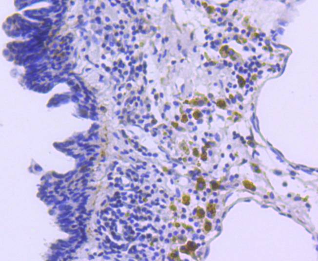
Immunohistochemical analysis of paraffin-embedded mouse lung tissue using anti-p27 KIP 1 antibody. Counter stained with hematoxylin.
Immunohistochemical analysis of paraffin-embedded mouse colon tissue using anti-p27 KIP 1 antibody. Counter stained with hematoxylin.
ICC staining p27 KIP 1 in A431 cells (green). The nuclear counter stain is DAPI (blue). Cells were fixed in paraformaldehyde, permeabilised with 0.25% Triton X100/PBS.
ICC staining p27 KIP 1 in MCF-7 cells (green). The nuclear counter stain is DAPI (blue). Cells were fixed in paraformaldehyde, permeabilised with 0.25% Triton X100/PBS.
Flow cytometric analysis of Hela cells with p27 KIP 1 antibody at 1/50 dilution (red) compared with an unlabelled control (cells without incubation with primary antibody; black). Alexa Fluor 488-conjugated goat anti rabbit IgG was used as the secondary antibody.
Cell cycle progression is regulated by a series of cyclin-dependent kinases consisting of catalytic subunits, designated Cdks, as well as activating subunits, designated cyclins. Orderly progression through the cell cycle requires the activation and inactivation of different cyclin-Cdks at appropriate times. A series of proteins has recently been described that function as "mitotic inhibitors." These include p21, the levels of which are elevated upon DNA damage in G1 in a p53-dependent manner; p16; and a more recently described p16-related inhibitor designated p15. A p21-related protein, p27, has been described as a negative regulator of G1 progression and speculated to function as a possible mediator of TGFβ-induced G1 arrest. p27 interacts strongly with D-type cyclins and Cdk4 in vitro and, to a lesser extent, with cyclin E and Cdk2.
If you have published an article using product 48842, please notify us so that we can cite your literature.


 Yes
Yes


