Product Detail
Product NameMCSF Rabbit mAb
Clone No.SU0413
Host SpeciesRecombinant Rabbit
Clonality Monoclonal
PurificationProA affinity purified
ApplicationsWB, IHC, ICC, IP
Species ReactivityHu
Immunogen Descrecombinant protein
ConjugateUnconjugated
Other NamesColony stimulating factor 1 (macrophage) antibody Colony stimulating factor 1 antibody Colony stimulating factor macrophage specific antibody CSF 1 antibody CSF-1 antibody CSF1 antibody CSF1_HUMAN antibody Csfm antibody Lanimostim antibody M CSF antibody M-CSF antibody Macrophage Colony Stimulating Factor 1 antibody Macrophage colony stimulating factor antibody MCSF antibody MGC31930 antibody Processed macrophage colony-stimulating factor 1 antibody
Accession NoSwiss-Prot#:P09603
Uniprot
P09603
Gene ID
1435;
Calculated MW60 kDa
Formulation1*TBS (pH7.4), 1%BSA, 40%Glycerol. Preservative: 0.05% Sodium Azide.
StorageStore at -20˚C
Application Details
WB: 1:500-1:1000
IHC: 1:50-1:100
ICC: 1:50-1:200
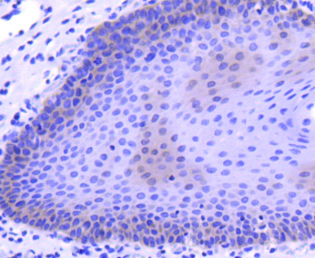
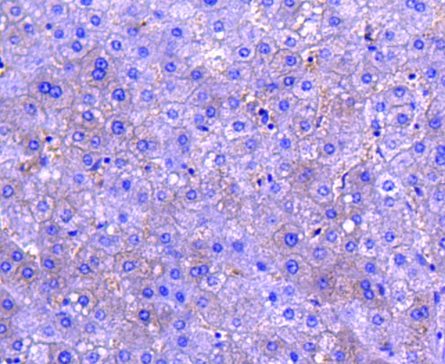
Immunohistochemical analysis of paraffin-embedded human tonsil tissue using anti-MCSF antibody. Counter stained with hematoxylin.
Immunohistochemical analysis of paraffin-embedded human lung tissue using anti-MCSF antibody. Counter stained with hematoxylin.
Immunohistochemical analysis of paraffin-embedded human liver tissue using anti-MCSF antibody. Counter stained with hematoxylin.
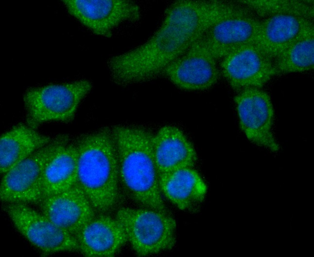
ICC staining MCSF in A431 cells (green). The nuclear counter stain is DAPI (blue). Cells were fixed in paraformaldehyde, permeabilised with 0.25% Triton X100/PBS.
ICC staining MCSF in HepG2 cells (green). The nuclear counter stain is DAPI (blue). Cells were fixed in paraformaldehyde, permeabilised with 0.25% Triton X100/PBS.
ICC staining MCSF in Hela cells (green). The nuclear counter stain is DAPI (blue). Cells were fixed in paraformaldehyde, permeabilised with 0.25% Triton X100/PBS.
The macrophage colony-stimulating factor (M-CSF), also designated CSF-1, was originally discovered in serum, urine and other biological fluids as a factor that can stimulate the formation of macrophage colonies from bone marrow hematopoietic progenitor cells. M-CSF is a homodimeric cytokine that is produced by fibroblasts, epithelial cells, bone marrow stromal cells, osteoblasts, keratinocytes, macrophages, T cells and B cells. M-CSF is a glycoprotein required for the proliferation and differentiation of mononuclear phagocytes, including osteoclasts. M-CSF has also been identified as an important mediator of the inflammatory response and can regulate the release of proinflammatory cytokines from macrophages. M-CSF exerts its pleiotropic effects by binding to a single type of high affinity cell surface receptor that is encoded by the c-Fms proto-oncogene.
If you have published an article using product 48850, please notify us so that we can cite your literature.


 Yes
Yes


