Product Detail
Product NameAndrogen receptor Rabbit mAb
Clone No.ST0453
Host SpeciesRecombinant Rabbit
Clonality Monoclonal
PurificationProA affinity purified
ApplicationsWB, ICC/IF, IHC, FC
Species ReactivityHu
Immunogen Descrecombinant protein
ConjugateUnconjugated
Other NamesAIS antibody ANDR_HUMAN antibody Androgen nuclear receptor variant 2 antibody Androgen receptor (dihydrotestosterone receptor; testicular feminization; spinal and bulbar muscular atrophy; Kennedy disease) antibody Androgen receptor antibody androgen receptor splice variant 4b antibody AR antibody AR8 antibody DHTR antibody Dihydro testosterone receptor antibody Dihydrotestosterone receptor (DHTR) antibody Dihydrotestosterone receptor antibody HUMARA antibody HYSP1 antibody KD antibody Kennedy disease (KD) antibody NR3C4 antibody Nuclear receptor subfamily 3 group C member 4 (NR3C4) antibody Nuclear receptor subfamily 3 group C member 4 antibody SBMA antibody SMAX1 antibody Spinal and bulbar muscular atrophy (SBMA) antibody Spinal and bulbar muscular atrophy antibody Testicular Feminization (TFM) antibody TFM antibody
Accession NoSwiss-Prot#:P10275
Uniprot
P10275
Gene ID
367;
Calculated MW99 kDa
Formulation1*TBS (pH7.4), 1%BSA, 40%Glycerol. Preservative: 0.05% Sodium Azide.
StorageStore at -20˚C
Application Details
WB: 1:500-1:1000
IHC: 1:50-1:100
ICC: 1:50-1:200
Immunohistochemical analysis of paraffin-embedded human breast carcinoma tissue using anti-Androgen receptor antibody. Counter stained with hematoxylin.
Immunohistochemical analysis of paraffin-embedded mouse testis tissue using anti-Androgen receptor antibody. Counter stained with hematoxylin.
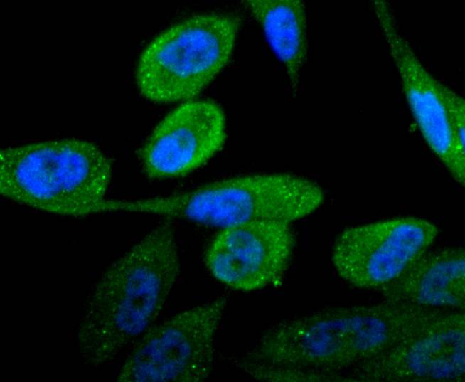
ICC staining Androgen receptor in PC-3M cells (green). The nuclear counter stain is DAPI (blue). Cells were fixed in paraformaldehyde, permeabilised with 0.25% Triton X100/PBS.
ICC staining Androgen receptor in N2A cells (green). The nuclear counter stain is DAPI (blue). Cells were fixed in paraformaldehyde, permeabilised with 0.25% Triton X100/PBS.
ICC staining Androgen receptor in MCF-7 cells (green). The nuclear counter stain is DAPI (blue). Cells were fixed in paraformaldehyde, permeabilised with 0.25% Triton X100/PBS.
Flow cytometric analysis of MCF-7 cells with Androgen receptor antibody at 1/50 dilution (red) compared with an unlabelled control (cells without incubation with primary antibody; black). Alexa Fluor 488-conjugated goat anti rabbit IgG was used as the secondary antibody
Androgens exhibit a wide range of effects on the development, maintenance and regulation of male phenotype and male reproductive physiology. The androgen receptor (AR) is a member of the steroid superfamily of ligand-dependent transcription factors. ARs bind the two biologically active androgens, testosterone (T) and dihydrotestosterone (DHT), with high and nearly identical affinities; however, the rates of association and dissociation of T are about three times more rapid than those of DHT. This difference has resulted in speculation as to whether these differences in binding kinetics could account for the different physiological effects of T and DHT. A striking feature of AR is its rapid degradation in the absence of ligand. It is now well established that androgen binding results in an at least six-fold increase in androgen stability and that ligand-induced stabilization of AR is highly androgen- specific.
If you have published an article using product 48858, please notify us so that we can cite your literature.


 Yes
Yes


