Product Detail
Product NameASH2L Rabbit mAb
Clone No.ST47-01
Host SpeciesRecombinant Rabbit
Clonality Monoclonal
PurificationProA affinity purified
ApplicationsWB, ICC/IF, IHC
Species ReactivityHu, Ms, Rt
Immunogen Descrecombinant protein
ConjugateUnconjugated
Other NamesASH 2 antibody Ash2 (absent small or homeotic) like antibody Ash2 (absent, small, or homeotic) like (Drosophila) antibody ASH2 antibody ASH2 LIKE antibody ASH2 like protein antibody ASH2-like protein antibody Ash2l antibody ASH2L_HUMAN antibody ASH2L1 antibody ASH2L2 antibody Bre 2 antibody Bre2 antibody Discs 2-like antibody Drosophila absent, small, or homeotic antibody Drosophila ASH2-like antibody Set1/Ash2 histone methyltransferase complex subunit ASH2 antibody
Accession NoSwiss-Prot#:Q9UBL3
Uniprot
Q9UBL3
Gene ID
9070;
Calculated MW69 kDa
Formulation1*TBS (pH7.4), 1%BSA, 40%Glycerol. Preservative: 0.05% Sodium Azide.
StorageStore at -20˚C
Application Details
WB: 1:1,000-5,000
IHC: 1:200-1:500
ICC: 1:200-1:500
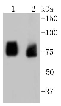
Western blot analysis of ASH2L on different lysates using anti-ASH2L antibody at 1/1,000 dilution. Positive control: Lane 1: PC12 Lane 2: 293T
Immunohistochemical analysis of paraffin-embedded mouse testis tissue using anti-ASH2L antibody. Counter stained with hematoxylin.
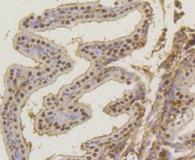
Immunohistochemical analysis of paraffin-embedded mouse colon tissue using anti-ASH2L antibody. Counter stained with hematoxylin.
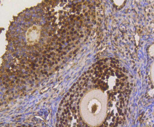
Immunohistochemical analysis of paraffin-embedded mouse ovary tissue using anti-ASH2L antibody. Counter stained with hematoxylin.
Immunohistochemical analysis of paraffin-embedded mouse placenta tissue using anti-ASH2L antibody. Counter stained with hematoxylin.
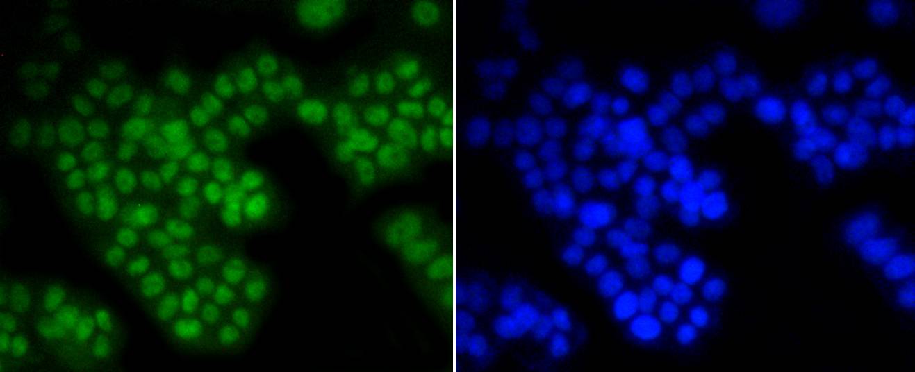
ICC staining ASH2L in PC12 cells (green). The nuclear counter stain is DAPI (blue). Cells were fixed in paraformaldehyde, permeabilised with 0.25% Triton X100/PBS.
ICC staining ASH2L in MCF-7 cells (green). The nuclear counter stain is DAPI (blue). Cells were fixed in paraformaldehyde, permeabilised with 0.25% Triton X100/PBS.
ICC staining ASH2L in CRC cells (green). Cells were fixed in paraformaldehyde, permeabilised with 0.25% Triton X100/PBS.
The human ASH2L gene encodes a 628 amino acid protein known as ASH2L1, or isoform 1, which contains a nuclear localization signal and PHD finger motif, suggesting that the gene product functions as a transcription regulator. Alternative splicing results in a shorter isoform 2, designated ASH2L2, which is missing the first 94 amino acid residues found in ASH2L1. Human ASH2L proteins are 60% homologous to Drosophila ash2, which positively regulates expression of certain genes in early development, and contain similar, but not identical, domains, including a zinc finger motif. ASH2L is highly expressed in fetal liver, testis and leukemia cell lines with erythroid and megakaryocytic potential, such as K562, Hel and Dami. Differentiation inducers (e.g. phorbol ester and hemin) cause different expression patterns in these cells lines, suggesting that ASH2L plays a role in hematopoiesis and is associated with particular types of leukemia.
If you have published an article using product 48863, please notify us so that we can cite your literature.


 Yes
Yes


