Product Detail
Product NameMAP1LC3A Rabbit mAb
Clone No.ST47-03
Host SpeciesRecombinant Rabbit
Clonality Monoclonal
PurificationProA affinity purified
ApplicationsWB, ICC/IF, IHC, IP, FC
Species ReactivityHu, Ms, Rt
Immunogen Descrecombinant protein
ConjugateUnconjugated
Other NamesATG8E antibody
Autophagy-related protein LC3 A antibody
Autophagy-related ubiquitin-like modifier LC3 A antibody
LC3 antibody
LC3A antibody
MAP1 light chain 3 like protein 1 antibody
MAP1 light chain 3-like protein 1 antibody
MAP1A/1B light chain 3 A antibody
MAP1A/MAP1B LC3 A antibody
MAP1A/MAP1B light chain 3 A antibody
MAP1ALC3 antibody
MAP1BLC3 antibody
Map1lc3a antibody
Microtubule associated proteins 1A/1B light chain 3 antibody
Microtubule-associated protein 1 light chain 3 alpha antibody
Microtubule-associated proteins 1A and 1B, light chain 3 antibody
Microtubule-associated proteins 1A/1B light chain 3A antibody
MLP3A_HUMAN antibody
Accession NoSwiss-Prot#:Q9H492
Uniprot
Q9H492
Gene ID
84557;
Calculated MW14 kDa
Formulation1*TBS (pH7.4), 1%BSA, 40%Glycerol. Preservative: 0.05% Sodium Azide.
StorageStore at -20˚C
Application Details
WB: 1:1,000-1:2,000
IHC: 1:50-1:200
ICC: 1:50-1:200
FC: 1:50-1:100
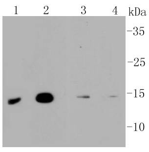
Western blot analysis of MAP1LC3A on different lysates using anti-MAP1LC3A antibody at 1/1,000 dilution. Positive control:
Lane 1: SHG-44
Lane 2: Mouse brain
Lane 3: Mouse liver
Lane 4: Mouse skeletal muscle
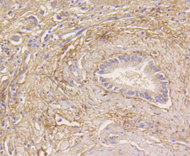
Immunohistochemical analysis of paraffin-embedded human liver tissue using anti-MAP1LC3A antibody. Counter stained with hematoxylin.
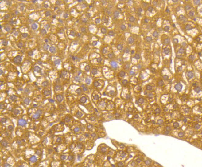
Immunohistochemical analysis of paraffin-embedded mouse liver tissue using anti-MAP1LC3A antibody. Counter stained with hematoxylin.
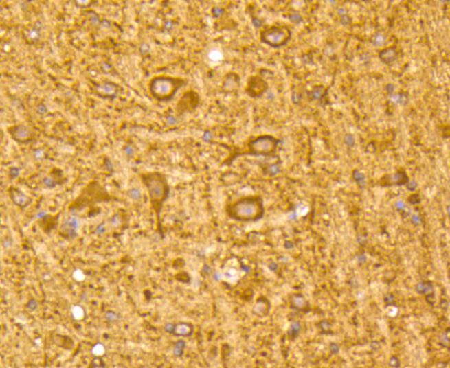
Immunohistochemical analysis of paraffin-embedded mouse brain tissue using anti-MAP1LC3A antibody. Counter stained with hematoxylin.
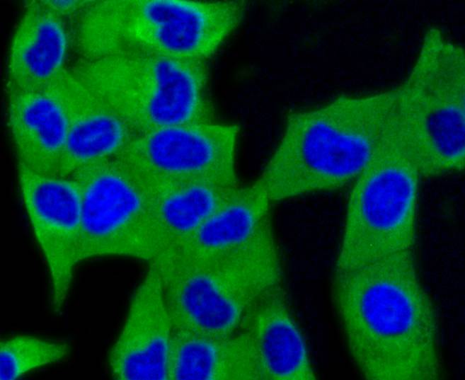
ICC staining MAP1LC3A in Hela cells (green). The nuclear counter stain is DAPI (blue). Cells were fixed in paraformaldehyde, permeabilised with 0.25% Triton X100/PBS.
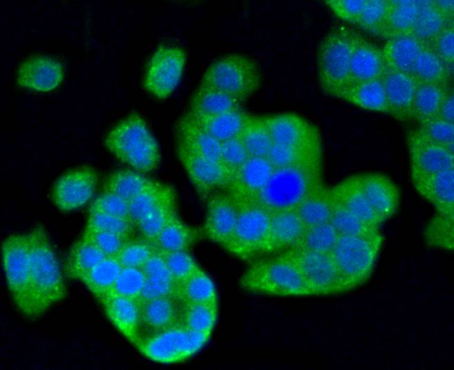
ICC staining MAP1LC3A in PC12 cells (green). The nuclear counter stain is DAPI (blue). Cells were fixed in paraformaldehyde, permeabilised with 0.25% Triton X100/PBS.
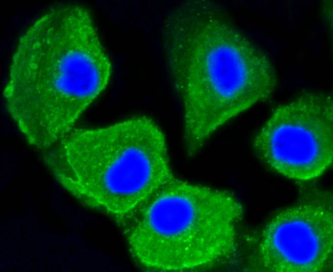
ICC staining MAP1LC3A in HUVEC cells (green). The nuclear counter stain is DAPI (blue). Cells were fixed in paraformaldehyde, permeabilised with 0.25% Triton X100/PBS.
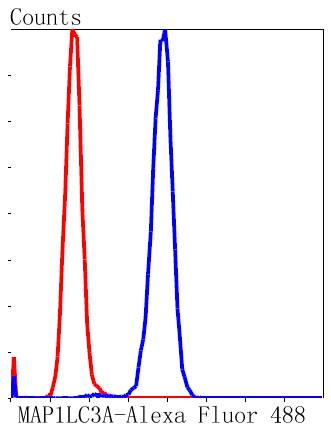
Flow cytometric analysis of SH-SY-5Y cells with MAP1LC3A antibody at 1/50 dilution (blue) compared with an unlabelled control (cells without incubation with primary antibody; red). Alexa Fluor 488-conjugated goat anti rabbit IgG was used as the secondary antibody.
Microtubules, the primary component of the cytoskeletal network, interact with proteins called microtubule-associated proteins (MAPs). The microtubule-associated proteins can be divided into two groups, structural and dynamic. The structural microtubule-associated proteins, MAP-1A, MAP-1B, MAP-2A, MAP-2B and MAP-2C, stimulate tubulin assembly, enhance micro-tubule stability and influence the spatial distribution of microtubules within cells. Both MAP-1 and, to a greater extent, MAP-2 have been implicated as agents of microtubule depolymerization by suppressing the dynamic instability of the microtubules. The suppression of microtubule dynamic instability by the MAP proteins is thought to be associated with phosphorylation of the MAPs.
If you have published an article using product 48865, please notify us so that we can cite your literature.


 Yes
Yes


