Product Detail
Product NameLysozyme Rabbit mAb
Clone No.ST50-02
Host SpeciesRecombinant Rabbit
Clonality Monoclonal
PurificationProA affinity purified
ApplicationsWB, ICC/IF, IHC, IP
Species ReactivityHu, Ms
Immunogen Descrecombinant protein
ConjugateUnconjugated
Other Names1 4 beta N acetylmuramidase C antibody 1 antibody 4-beta-N-acetylmuramidase C antibody EC 3.2.1.17 antibody LYSC_HUMAN antibody Lysosyme antibody Lysozyme (renal amyloidosis) antibody Lysozyme C antibody Lysozyme C precursor antibody LYZ antibody LZM antibody Renal amyloidosis antibody
Accession NoSwiss-Prot#:P61626
Uniprot
P61626
Gene ID
4069;
Calculated MW17 kDa
Formulation1*TBS (pH7.4), 1%BSA, 40%Glycerol. Preservative: 0.05% Sodium Azide.
StorageStore at -20˚C
Application Details
WB: 1:1,000-1:2,000
IHC: 1:200-1:500
ICC: 1:50-1:200
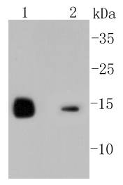
Western blot analysis of Lysozyme on different lysates using anti-Lysozyme antibody at 1/1,000 dilution. Positive control: Lane 1: Mouse kidney Lane 2: HL-60
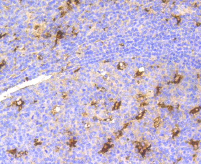
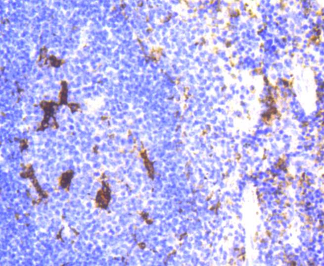
Immunohistochemical analysis of paraffin-embedded human tonsil tissue using anti-Lysozyme antibody. Counter stained with hematoxylin.
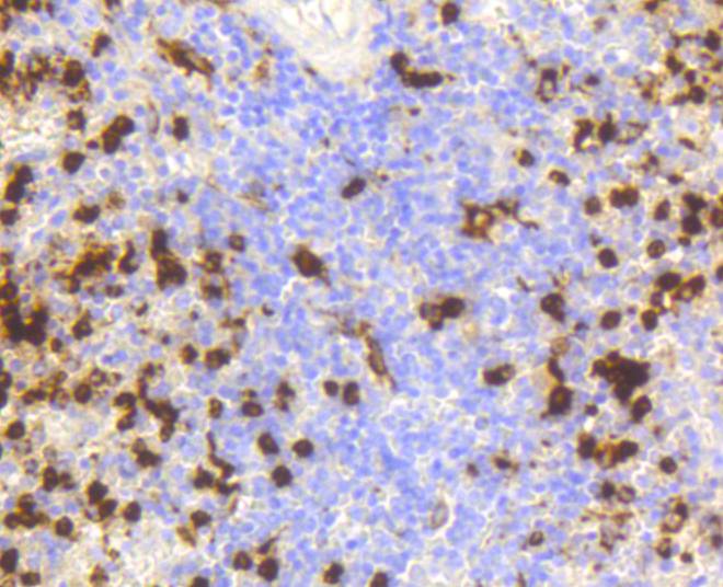
Immunohistochemical analysis of paraffin-embedded human spleen tissue using anti-Lysozyme antibody. Counter stained with hematoxylin. The nuclear counter stain is DAPI (blue).
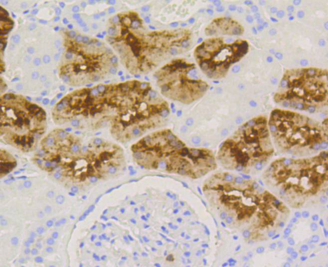
Immunohistochemical analysis of paraffin-embedded human kidney tissue using anti-Lysozyme antibody. Counter stained with hematoxylin.
Immunohistochemical analysis of paraffin-embedded mouse spleen tissue using anti-Lysozyme antibody. Counter stained with hematoxylin.
Immunohistochemical analysis of paraffin-embedded mouse kidney tissue using anti-Lysozyme antibody. Counter stained with hematoxylin.
ICC staining Lysozyme in CRC cells (green). The nuclear counter stain is DAPI (blue). Cells were fixed in paraformaldehyde, permeabilised with 0.25% Triton X100/PBS.
The origins of the lysozyme proteins date back an estimated 400 to 600 million years. Generally, lysozyme genes are relatively small, roughly 10 kilobases in length, and composed of four exons and three introns. Originally a bacteriolytic defensive agent, the function of this family of proteins adapted to serve a digestive function in its present forms. Lysozymes in tissues and body fluids are associated with the monocyte-macrophage system and enhance the activity of immunoagents. Lysozyme C belongs to the glycosyl hydrolase 22 family, and newly identified relatives of Lysozyme C appear to possess anti-HIV activity, as well as preserved bacteriolytic function against Micrococcus lysodeikticus. Lysozyme C is capable of both hydrolysis and transglycosylation and also a slight esterase activity. It acts rapidly on both peptide-substituted and unsubstituted peptidoglycan, and slowly on chitin oligosaccharides. Lysozyme C defects are a cause of amyloidosis VIII, also called familial visceral or Ostertag-type amyloidosis.
If you have published an article using product 48872, please notify us so that we can cite your literature.




 Yes
Yes


