Product Detail
Product Namebeta Arrestin 1 Rabbit mAb
Clone No.ST51-08
Host SpeciesRecombinant Rabbit
Clonality Monoclonal
PurificationProA affinity purified
ApplicationsWB, ICC/IF, IHC, IP, FC
Species ReactivityHu, Ms, Rt
Immunogen Descrecombinant protein
ConjugateUnconjugated
Other NamesARB1 antibody
ARR1 antibody
ARRB1 antibody
ARRB1_HUMAN antibody
Arrestin 2 antibody
Arrestin beta 1 antibody
Arrestin beta-1 antibody
Beta-arrestin-1 antibody
Accession NoSwiss-Prot#:P49407
Uniprot
P49407
Gene ID
408;
Calculated MW50 kDa
Formulation1*TBS (pH7.4), 1%BSA, 40%Glycerol. Preservative: 0.05% Sodium Azide.
StorageStore at -20˚C
Application Details
WB: 1:1,000-1:2,000
IHC: 1:50-1:100
ICC: 1:50-1:200
FC: 1:50-1:100
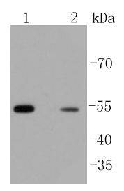
Western blot analysis of beta Arrestin 1 on different lysates using anti-beta Arrestin 1 antibody at 1/1,000 dilution. Positive control:
Lane 1: PC12
Lane 2: Jurkat
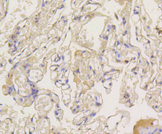
Immunohistochemical analysis of paraffin-embedded human lung tissue using anti-beta Arrestin 1 antibody. Counter stained with hematoxylin.
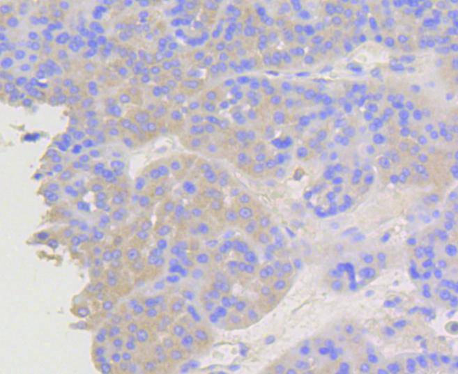
Immunohistochemical analysis of paraffin-embedded human liver cancer tissue using anti-beta Arrestin 1 antibody. Counter stained with hematoxylin.
Immunohistochemical analysis of paraffin-embedded human breast carcinoma tissue using anti-beta Arrestin 1 antibody. Counter stained with hematoxylin.
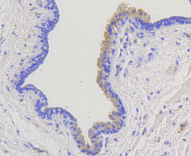
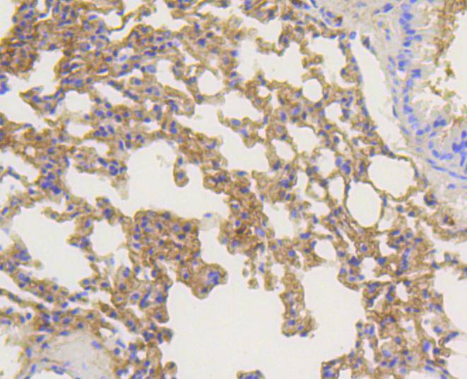
Immunohistochemical analysis of paraffin-embedded mouse lung tissue using anti-beta Arrestin 1 antibody. Counter stained with hematoxylin.
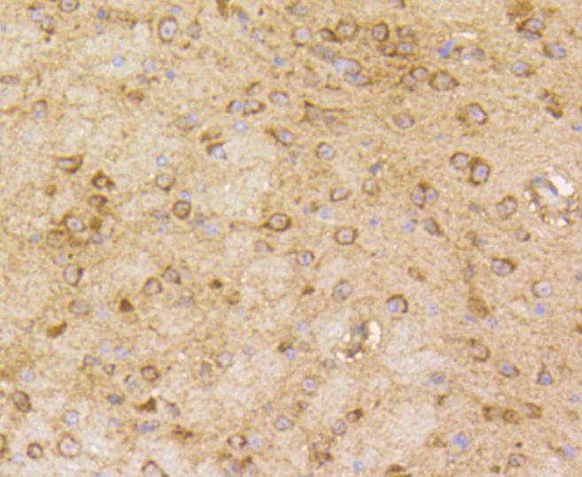
Immunohistochemical analysis of paraffin-embedded mouse brain tissue using anti-beta Arrestin 1 antibody. Counter stained with hematoxylin.
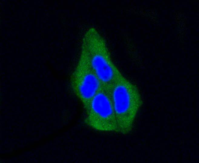
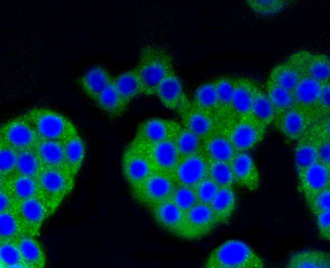
ICC staining beta Arrestin 1 in Hela cells (green). The nuclear counter stain is DAPI (blue). Cells were fixed in paraformaldehyde, permeabilised with 0.25% Triton X100/PBS.
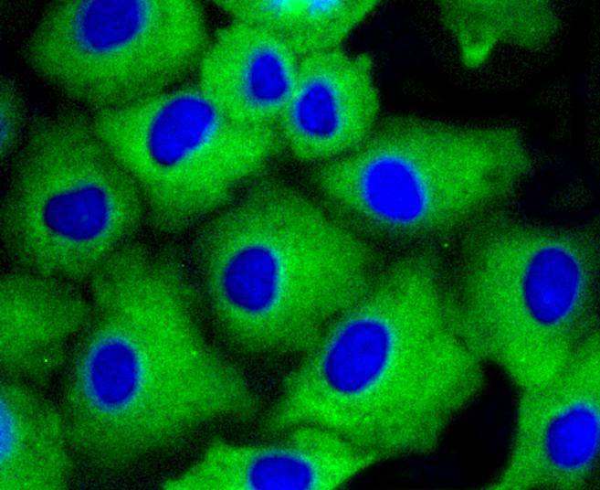
ICC staining beta Arrestin 1 in A549 cells (green). The nuclear counter stain is DAPI (blue). Cells were fixed in paraformaldehyde, permeabilised with 0.25% Triton X100/PBS.
ICC staining beta Arrestin 1 in PC12 cells (green). The nuclear counter stain is DAPI (blue). Cells were fixed in paraformaldehyde, permeabilised with 0.25% Triton X100/PBS.
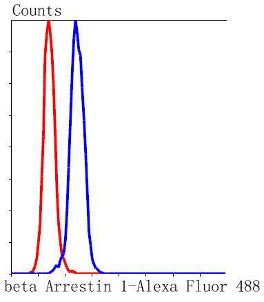
Flow cytometric analysis of Hela cells with beta Arrestin 1 antibody at 1/50 dilution (blue) compared with an unlabelled control (cells without incubation with primary antibody; red). Alexa Fluor 488-conjugated goat anti rabbit IgG was used as the secondary antibody.
The members of the G protein coupled receptor family are distinguished by their slow transmitting response to ligand binding. These seven transmembrane proteins include the adrenergic, serotonin and dopamine receptors. The effect of the signaling molecule can be excitatory or inhibitory depending on the type of receptor to which it binds. Members of theβ-Arrestin family regulate receptor binding to G proteins. β-Arrestins have been found to be located at postsynaptic sites, where they are thought to act in concert withβARK (βARK1, also designated GRK 2, orβARK2, also designated GRK 3 to regulate G protein-coupled neurotransmitter receptors. Expression ofβ-Arrestin-1 and b-Arrestin-2 is seen predominantly in spleen and neuronal tissues. It has been shown thatβ-Arrestin-1 expression is modulated by intracellular cAMP, which may be a novel mechanism for the regulation of receptor-mediated responses.
If you have published an article using product 48875, please notify us so that we can cite your literature.


 Yes
Yes


