Product Detail
Product NameICAM1 Rabbit mAb
Clone No.ST0487
Host SpeciesRecombinant Rabbit
Clonality Monoclonal
PurificationProA affinity purified
ApplicationsWB, ICC/IF, IHC
Species ReactivityHu
Immunogen Descrecombinant protein
ConjugateUnconjugated
Other NamesAntigen identified by monoclonal antibody BB2 antibody BB 2 antibody BB2 antibody CD 54 antibody CD_antigen=CD54 antibody CD54 antibody Cell surface glycoprotein P3.58 antibody Human rhinovirus receptor antibody ICAM 1 antibody ICAM-1 antibody ICAM1 antibody ICAM1_HUMAN antibody intercellular adhesion molecule 1 (CD54), human rhinovirus receptor antibody Intercellular adhesion molecule 1 antibody Major group rhinovirus receptor antibody MALA 2 antibody MALA2 antibody MyD 10 antibody MyD10 antibody P3.58 antibody Surface antigen of activated B cells, BB2 antibody
Accession NoSwiss-Prot#:P05362
Uniprot
P05362
Gene ID
3383;
Calculated MW89 kDa
Formulation1*TBS (pH7.4), 1%BSA, 40%Glycerol. Preservative: 0.05% Sodium Azide.
StorageStore at -20˚C
Application Details
WB: 1:1,000-5,000
IHC: 1:50-1:200
ICC: 1:50-1:200
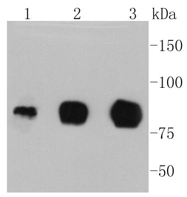
Western blot analysis of ICAM1 on different lysates using anti-ICAM1 antibody at 1/1,000 dilution. Positive control: Lane 1: Hela Lane 2: HUVEC Lane 3: Raji
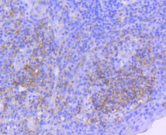
Immunohistochemical analysis of paraffin-embedded human tonsil tissue using anti-ICAM1 antibody. Counter stained with hematoxylin.
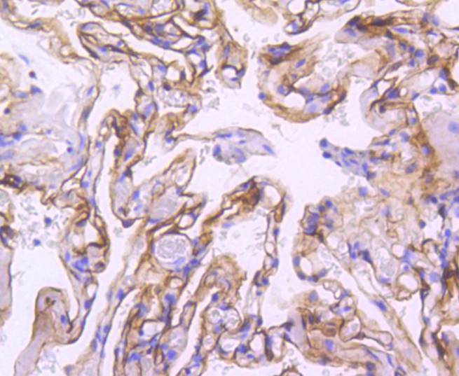
Immunohistochemical analysis of paraffin-embedded human lung tissue using anti-ICAM1 antibody. Counter stained with hematoxylin.
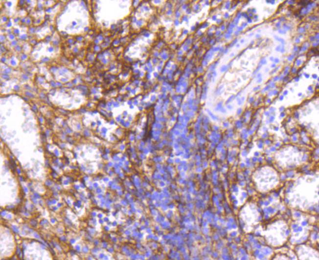
Immunohistochemical analysis of paraffin-embedded human spleen tissue using anti-ICAM1 antibody. Counter stained with hematoxylin.
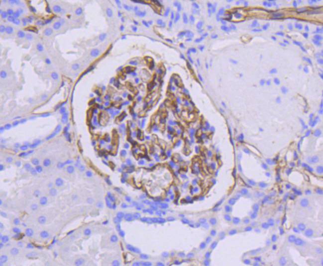
Immunohistochemical analysis of paraffin-embedded human kidney tissue using anti-ICAM1 antibody. Counter stained with hematoxylin.
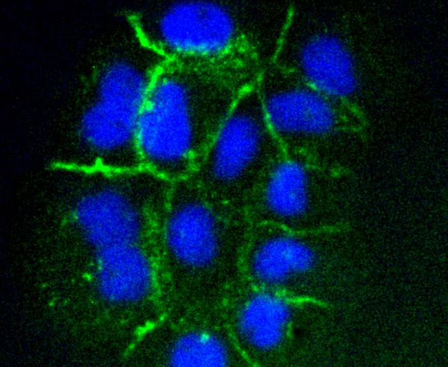
ICC staining ICAM1 in A431 cells (green). The nuclear counter stain is DAPI (blue). Cells were fixed in paraformaldehyde, permeabilised with 0.25% Triton X100/PBS.
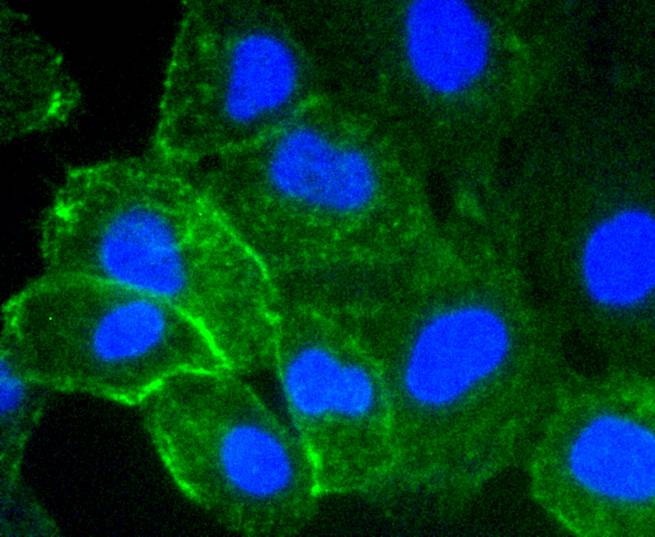
ICC staining ICAM1 in HUVEC cells (green). The nuclear counter stain is DAPI (blue). Cells were fixed in paraformaldehyde, permeabilised with 0.25% Triton X100/PBS.
Cell adhesion molecules (CAMs) are a family of closely related cell surface glycoproteins involved in cell-cell interactions during growth and are thought to play important, yet separate, roles in embryogenesis and development. The intracellular adhesion molecule-1 (ICAM-1), also referred to as CD54, is an integral membrane protein of the immunoglobulin superfamily and recognizes the beta2alpha1 and beta2alphaM Integrins. ICAM-2 functions as a ligand for lymphocyte function-associated antigen-1 (LFA-1) and is involved in leukocyte adhesion. ICAM-3 is highly expressed on the surface of human eosinophils and, when bound to ligand, may inhibit eosinophil inflammatory responses and survival. ICAM-4, also known as LW glycoprotein, interacts with Integrins alphaLbeta2, alphaMbeta2, alpha4beta1, the alphaV family and alphaIIbbeta3, and selective binding to different integrins may be relevant to the pathology in a number of red blood cell associated diseases. Lastly, ICAM-5, expressed on telencephalic neurons, binds CD11a/CD18 and thus may act as an adhesion molecule for leukocyte binding in the central nervous system.
If you have published an article using product 48883, please notify us so that we can cite your literature.


 Yes
Yes


