Product Detail
Product NamePBP Rabbit mAb
Clone No.SC58-09
Host SpeciesRecombinant Rabbit
Clonality Monoclonal
PurificationProA affinity purified
ApplicationsWB, ICC/IF, IHC, IP
Species ReactivityHu, Ms, Rt, zebrafish
Immunogen Descrecombinant protein
ConjugateUnconjugated
Other NamesEpididymis luminal protein 210 antibody Epididymis secretory protein Li 34 antibody Epididymis secretory protein Li 96 antibody HCNP antibody HCNPpp antibody HEL 210 antibody HEL S 34 antibody HEL S 96 antibody Hippocampal cholinergic neurostimulating peptide antibody Neuropolypeptide h3 antibody PBP antibody PEBP antibody PEBP 1 antibody PEBP-1 antibody Pebp1 antibody PEBP1_HUMAN antibody Phosphatidylethanolamine binding protein antibody Phosphatidylethanolamine binding protein 1 antibody Prostatic binding protein antibody Prostatic-binding protein antibody R kip antibody Raf kinase inhibitor protein antibody Raf kinase inhibitory protein antibody RKIP antibody
Accession NoSwiss-Prot#:P30086
Uniprot
P30086
Gene ID
5037;
Calculated MW21 kDa
Formulation1*TBS (pH7.4), 1%BSA, 40%Glycerol. Preservative: 0.05% Sodium Azide.
StorageStore at -20˚C
Application Details
WB: 1:1,000-5,000
IHC: 1:50-1:200
ICC: 1:50-1:200
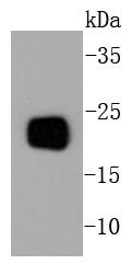
Western blot analysis of PBP on mouse brain lysates using anti-PBP antibody at 1/1,000 dilution.
Immunohistochemical analysis of paraffin-embedded human kidney tissue using anti-PBP antibody. Counter stained with hematoxylin.
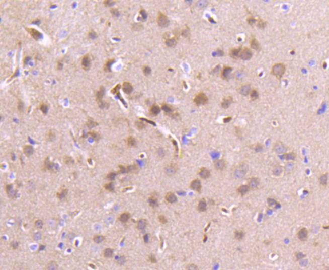
Immunohistochemical analysis of paraffin-embedded mouse brain tissue using anti-PBP antibody. Counter stained with hematoxylin.
Immunohistochemical analysis of paraffin-embedded mouse kidney tissue using anti-PBP antibody. Counter stained with hematoxylin.
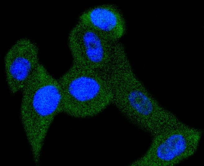
ICC staining PBP in PC-3M cells (green). The nuclear counter stain is DAPI (blue). Cells were fixed in paraformaldehyde, permeabilised with 0.25% Triton X100/PBS.
Members of the a-chemokine subfamily of inducible, secreted, pro-inflammatory cytokines contain a similar motif, in which the first two cysteine residues are separated by a single residue (Cys-X-Cys), and are also chemotactic for neutrophils. The platelet basic protein (PBP), a member of the a(lpha)-chemokine family, resides in the a(lpha)-granules of platelets and is released upon their activation. Proteolytic cleavage of the amino terminus of PBP leads to the generation of several peptides, which include mature PBP, connective tissue-activating peptide III (CTAP III, also designated low affinity platelet factor IV (LA-PF4)), b-thromboglobulin (b-TG), and neutrophil-activating peptide 2 (NAP-2). PBP and its N-truncated derivatives mediate inflammation and wound healing. Specifically, NAP-2 activates chemotaxis and degranulation in neutrophils during inflammation. The gene encoding human PBP maps to chromosome 4q12-q13.
If you have published an article using product 48929, please notify us so that we can cite your literature.


 Yes
Yes


