Product Detail
Product NamePrion Protein(PrP) Rabbit mAb
Clone No.SC57-05
Host SpeciesRecombinant Rabbit
ClonalityMonoclonal
PurificationProA affinity purified
ApplicationsWB, ICC/IF, IHC, FC
Species ReactivityHu, Ms, Rt
Immunogen Descrecombinant protein
ConjugateUnconjugated
Other NamesAlternative prion protein; major prion protein antibody AltPrP antibody ASCR antibody CD230 antibody CD230 antigen antibody CJD antibody GSS antibody KURU antibody Major prion protein antibody p27 30 antibody PRIO_HUMAN antibody Prion protein antibody Prion related protein antibody PRIP antibody PRNP antibody PrP antibody PrP27 30 antibody PrP27-30 antibody PrP33-35C antibody PrPC antibody PrPSc antibody Sinc antibody
Accession NoSwiss-Prot#:P04156
Uniprot
P04156
Gene ID
5621;
Calculated MW28 kDa
Formulation1*TBS (pH7.4), 1%BSA, 40%Glycerol. Preservative: 0.05% Sodium Azide.
StorageStore at -20˚C
Application Details
WB: 1:1,000-5,000
IHC: 1:50-1:200
ICC: 1:50-1:200
FC: 1:50-1:100
Western blot analysis of PrP on different lysates using anti-PrP antibody at 1/1,000 dilution. Positive control: Lane 1: Rat brain Lane 2: Mouse brain
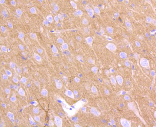
Immunohistochemical analysis of paraffin-embedded rat brain tissue using anti-PrP antibody. Counter stained with hematoxylin.
Immunohistochemical analysis of paraffin-embedded mouse brain tissue using anti-PrP antibody. Counter stained with hematoxylin.
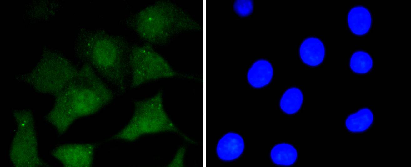
ICC staining PrP in N2A cells (green). The nuclear counter stain is DAPI (blue). Cells were fixed in paraformaldehyde, permeabilised with 0.25% Triton X100/PBS.
ICC staining PrP in SHG-44 cells (green). The nuclear counter stain is DAPI (blue). Cells were fixed in paraformaldehyde, permeabilised with 0.25% Triton X100/PBS.
ICC staining PrP in SH-SY-5Y cells (green). The nuclear counter stain is DAPI (blue). Cells were fixed in paraformaldehyde, permeabilised with 0.25% Triton X100/PBS.
Flow cytometric analysis of SH-SY-5Y cells with PrP antibody at 1/50 dilution (red) compared with an unlabelled control (cells without incubation with primary antibody; black). Alexa Fluor 488-conjugated goat anti rabbit IgG was used as the secondary antibody.
Prion diseases, or transmissible spongiform encephalopathies (TSEs), are manifested as genetic, infectious or sporadic, lethal neurodegenerative disorders involving alterations of the prion protein (PrP). Characteristic of prion diseases, cellular PrP (PrPc) is converted to the disease form, PrPSc, through alterations in the protein folding conformations. PrPc is constitutively expressed in normal adult brain and is sensitive to proteinase K digestion, while the altered PrPSc conformation is resistant to proteases, resulting in a distinct molecular mass after PK treatment. Consistent with the transient infection process of prion diseases, incubation of PrPc with PrPSc both in vitro and in vivo produces PrPc that is resistant to protease degradation. Infectious PrPSc is found at high levels in the brains of animals affected by TSEs, including scrapie in sheep, BSE in cattle and Cruetzfeldt-Jakob disease in humans.
If you have published an article using product 48939, please notify us so that we can cite your literature.


 Yes
Yes


