Product Detail
Product NameKu80 Rabbit mAb
Clone No.SC06-14
Host SpeciesRecombinant Rabbit
Clonality Monoclonal
PurificationProA affinity purified
ApplicationsWB, ICC/IF, IHC, IP
Species ReactivityHu
Immunogen Descrecombinant protein
ConjugateUnconjugated
Other Names86 kDa subunit of Ku antigen antibody ATP dependent DNA helicase 2 subunit 2 antibody ATP dependent DNA helicase II 80 kDa subunit antibody ATP dependent DNA helicase II 86 Kd subunit antibody ATP dependent DNA helicase II antibody ATP-dependent DNA helicase 2 subunit 2 antibody ATP-dependent DNA helicase II 80 kDa subunit antibody CTC box binding factor 85 kDa antibody CTC box-binding factor 85 kDa subunit antibody CTC85 antibody CTCBF antibody DNA repair protein XRCC5 antibody KARP 1 antibody KARP1 antibody Ku 80 antibody Ku autoantigen 80kDa antibody Ku80 antibody Ku86 antibody Ku86 autoantigen related protein 1 antibody KUB 2 antibody KUB2 antibody Lupus Ku autoantigen protein p86 antibody NFIV antibody Nuclear factor IV antibody Thyroid lupus autoantigen antibody Thyroid-lupus autoantigen antibody TLAA antibody X ray repair complementing defective repair in Chinese hamster cells 5 (double strand break rejoining) antibody X-ray repair complementing defective repair in Chinese hamster cells 5 (double-strand-break rejoining) antibody X-ray repair cross-complementing protein 5 antibody Xray repair complementing defective repair in Chinese hamster cells 5 antibody XRCC 5 antibody XRCC5 antibody XRCC5_HUMAN antibody
Accession NoSwiss-Prot#:P13010
Uniprot
P13010
Gene ID
7520;
Calculated MW83 kDa
Formulation1*TBS (pH7.4), 1%BSA, 40%Glycerol. Preservative: 0.05% Sodium Azide.
StorageStore at -20˚C
Application Details
WB: 1:1,000-1:2,000
IHC: 1:50-1:200
ICC: 1:50-1:200
Western blot analysis of Ku80 on MCF-7 cells lysates using anti-Ku80 antibody at 1/1,000 dilution.
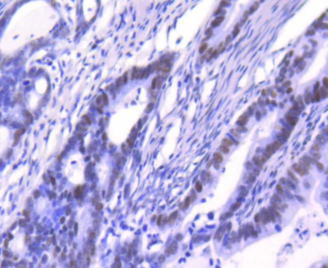
Immunohistochemical analysis of paraffin-embedded human colon cancer tissue using anti-Ku80 antibody. Counter stained with hematoxylin.
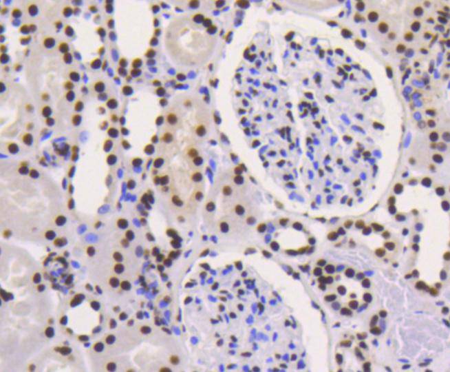
Immunohistochemical analysis of paraffin-embedded human kideny tissue using anti-Ku80 antibody. Counter stained with hematoxylin.
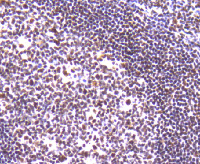
Immunohistochemical analysis of paraffin-embedded human tonsil tissue using anti-Ku80 antibody. Counter stained with hematoxylin.
Immunohistochemical analysis of paraffin-embedded human breast carcinoma tissue using anti-Ku80 antibody. Counter stained with hematoxylin.
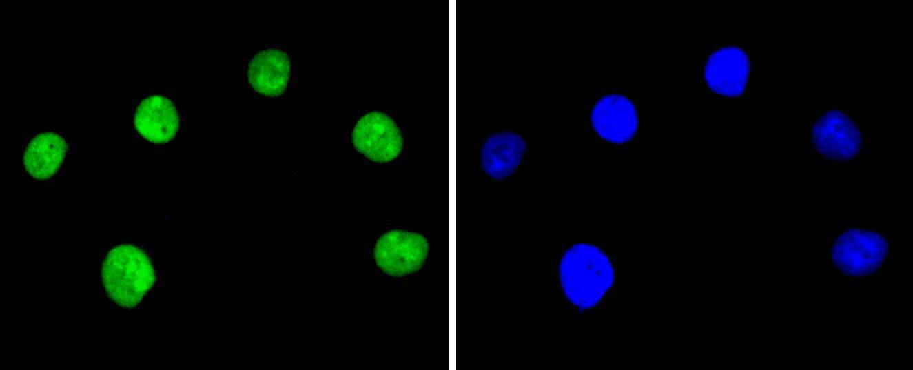
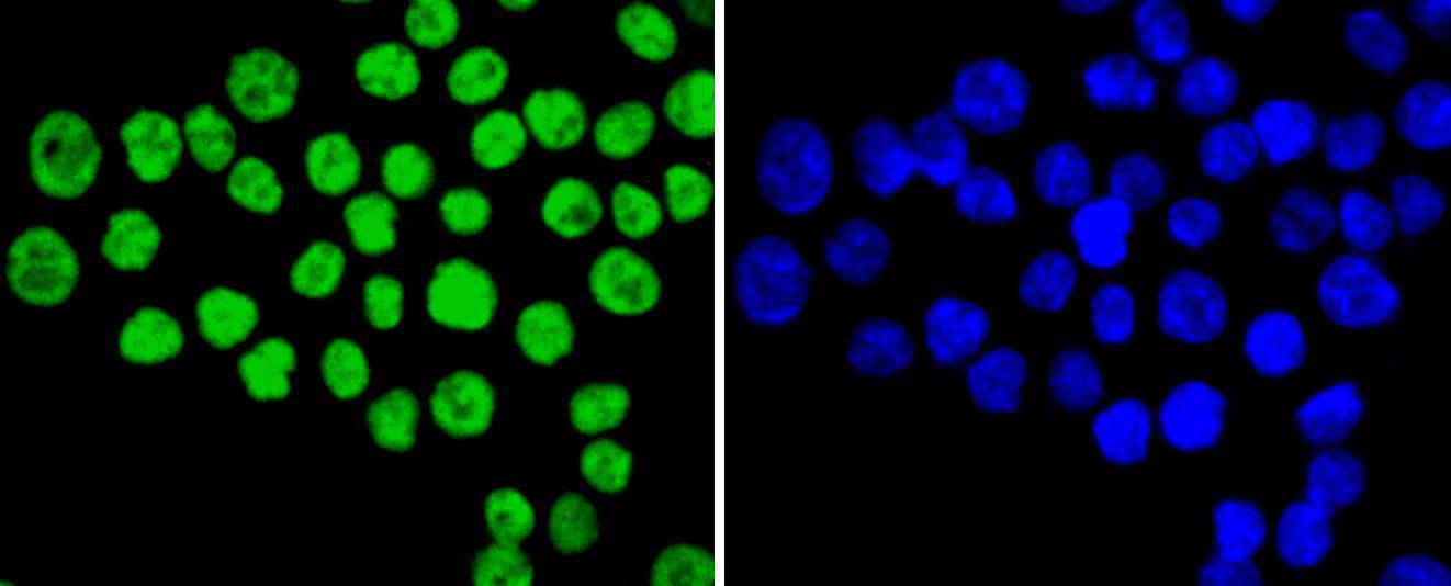
ICC staining Ku80 in Hela cells (green). The nuclear counter stain is DAPI (blue). Cells were fixed in paraformaldehyde, permeabilised with 0.25% Triton X100/PBS.
ICC staining Ku80 in A549 cells (green). The nuclear counter stain is DAPI (blue). Cells were fixed in paraformaldehyde, permeabilised with 0.25% Triton X100/PBS.
ICC staining Ku80 in SW480 cells (green). The nuclear counter stain is DAPI (blue). Cells were fixed in paraformaldehyde, permeabilised with 0.25% Triton X100/PBS.
The Ku protein is localized in the nucleus and is composed of subunits referred to as Ku-70 (p70) and Ku-86 (p86) which is also known by the synonym Ku-80 or (p80). Ku was first described as an autoantigen to which antibodies were produced in a patient with scleroderma polymyositis overlap syndrome, and was later found in the sera of patients with other rheumatic diseases. Both subunits of the Ku protein have been cloned, and a number of functions have been proposed for Ku, including cell signaling, DNA replication and transcriptional activation. Ku is involved in Pol II-directed transcription by virtue of its DNA binding activity, serving as the regulatory component of the DNA-associated protein kinase that phosphorylates Pol II and transcription factor Sp. Ku proteins also activate transcription from the U1 small nuclear RNA and the human transferrin receptor gene promoters. A Ku-related protein designated the enhancer 1 binding factor (E1BF), composed of two subunits, has been identified as a positive regulator of RNA polymerase I transcription initiation.
If you have published an article using product 48954, please notify us so that we can cite your literature.




 Yes
Yes


