Product Detail
Product NameTopoisomerase I Rabbit mAb
Clone No.SC69-03
Host SpeciesRecombinant Rabbit
Clonality Monoclonal
PurificationProA affinity purified
ApplicationsWB, ICC/IF, IHC, FC
Species ReactivityHu, Ms
Immunogen Descrecombinant protein
ConjugateUnconjugated
Other NamesDNA topoisomerase 1 antibody DNA topoisomerase I antibody NUP98 fusion gene antibody TOP 1 antibody TOP I antibody TOP1 antibody TOP1_HUMAN antibody TOPI antibody Topoisomerase (DNA) I antibody Topoisomerase 1 antibody Topoisomerase1 antibody TopoisomeraseI antibody Type I DNA topoisomerase antibody
Accession NoSwiss-Prot#:P11387
Uniprot
P11387
Gene ID
7150;
Calculated MW91 kDa
Formulation1*TBS (pH7.4), 1%BSA, 40%Glycerol. Preservative: 0.05% Sodium Azide.
StorageStore at -20˚C
Application Details
WB: 1:1,000-5,000
IHC: 1:50-1:200
ICC: 1:50-1:500
FC: 1:50-1:100
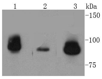
Western blot analysis of TOP1 on different lysates using anti-TOP1 antibody at 1/1,000 dilution. Positive control: Lane 1: HepG2 Lane 2: Jurkat Lane 3: MCF-7
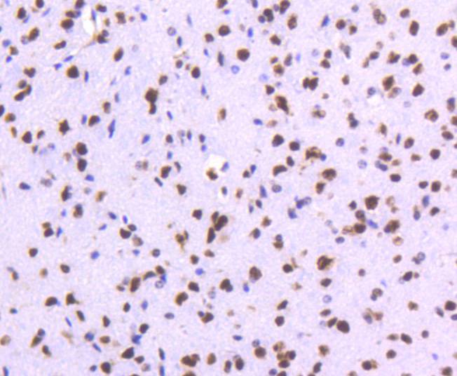
Immunohistochemical analysis of paraffin-embedded mouse brain tissue using anti-TOP1 antibody. Counter stained with hematoxylin.
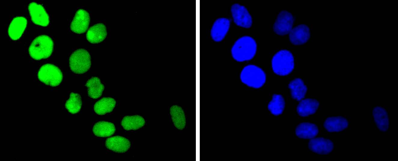
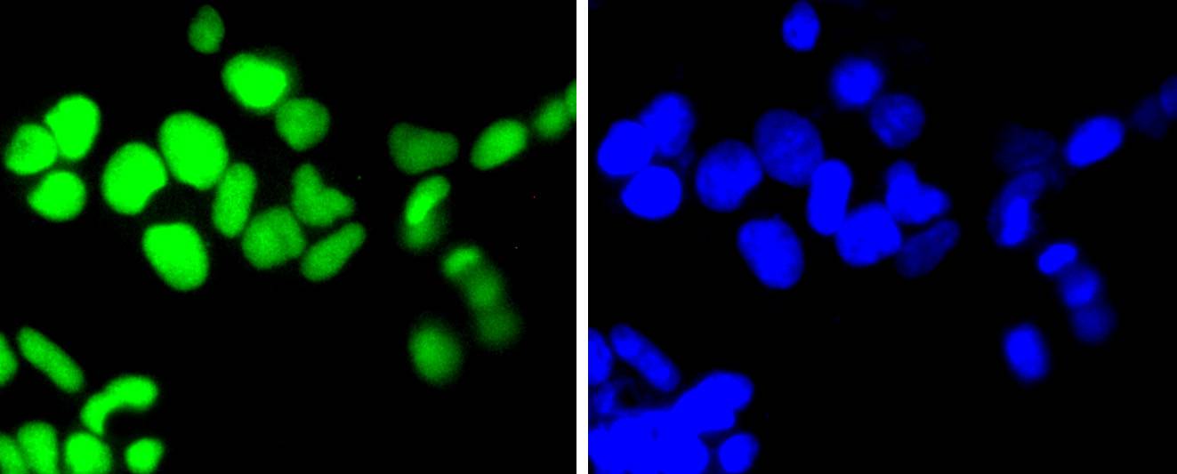
ICC staining TOP1 in Hela cells (green). The nuclear counter stain is DAPI (blue). Cells were fixed in paraformaldehyde, permeabilised with 0.25% Triton X100/PBS.
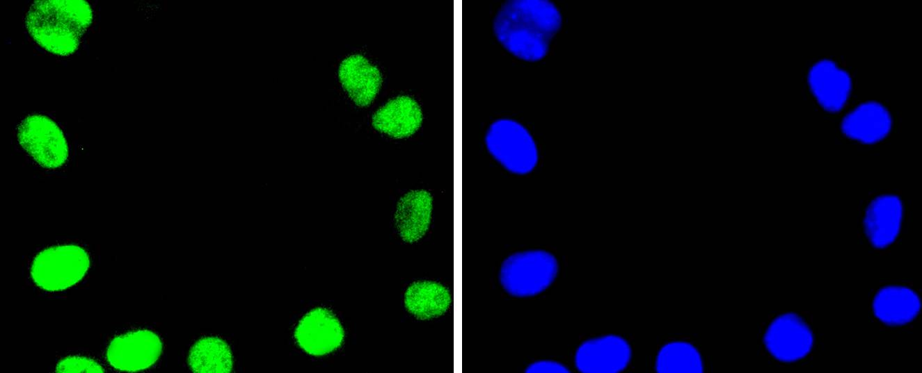
ICC staining TOP1 in SHG-44 cells (green). The nuclear counter stain is DAPI (blue). Cells were fixed in paraformaldehyde, permeabilised with 0.25% Triton X100/PBS.
ICC staining TOP1 in 293 cells (green). The nuclear counter stain is DAPI (blue). Cells were fixed in paraformaldehyde, permeabilised with 0.25% Triton X100/PBS.
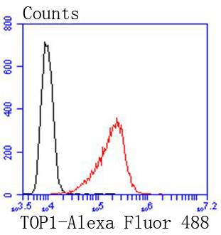
Flow cytometric analysis of HepG2 cells with TOP1 antibody at 1/50 dilution (red) compared with an unlabelled control (cells without incubation with primary antibody; black). Alexa Fluor 488-conjugated goat anti rabbit IgG was used as the secondary antibody
DNA topoisomerases play essential roles in many DNA metabolic processes including DNA repair. Topoisomerases can introduce DNA damage upon exposure to drugs that stabilize the covalent protein-DNA intermediate of the topoisomerase reaction. Lesions in DNA are also able to trap topoisomerase-DNA intermediates. DNA topoisomerase I (Top1) catalyzes the relaxation of supercoiled DNA by a mechanism of transient DNA strand cleavage characterized by the formation of a phosphotyrosyl bond between the DNA end and active site tyrosine. The antitumor agent camptothecin (CPT) reversibly stabilizes the covalent enzyme-DNA intermediate by inhibiting DNA religation. When a replication fork collides with a DNA Top1 cleavage complex, the covalently bound enzyme must be removed from the DNA 3' end before recombination-dependent replication restart. The tyrosyl-DNA phosphodiesterase Tdp1 and the structure-specific endonuclease Rad1-Rad10 function as primary alternative pathways of Top1 repair in Saccharomyces cerevisiae. In the budding yeast S. cerevisiae, DNA topoisomerases I and II can functionally substitute for each other in removing positive and negative DNA supercoils.
If you have published an article using product 48978, please notify us so that we can cite your literature.


 Yes
Yes


