Product Detail
Product NameMast Cell Tryptase Rabbit mAb
Clone No.SC68-07
Host SpeciesRecombinant Rabbit
Clonality Monoclonal
PurificationProA affinity purified
ApplicationsWB, IHC, IP
Species ReactivityHu, Ms, Rt
Immunogen Descrecombinant protein
ConjugateUnconjugated
Other Namesalpha II antibody Lung tryptase antibody Mast cell alpha II tryptase antibody Mast cell beta I tryptase antibody Mast cell protease 7 antibody Mast cell protease II antibody MCP 7 antibody Pituitary tryptase antibody Skin tryptase antibody TPS 1 antibody TPS1 antibody TPS2 antibody TPSAB1 antibody TPSAB1 protein antibody TPSB1 antibody Tryptase 1 antibody Tryptase alpha 1 antibody tryptase alpha I included antibody Tryptase alpha II antibody tryptase alpha II included antibody tryptase alpha included antibody tryptase alpha/beta 1 antibody Tryptase beta 1 antibody tryptase beta I included antibody Tryptase I antibody tryptase I included antibody Tryptase III antibody Tryptase skin antibody
Accession NoSwiss-Prot#:Q15661
Uniprot
Q15661
Gene ID
7177;
Calculated MW30 kDa
Formulation1*TBS (pH7.4), 1%BSA, 40%Glycerol. Preservative: 0.05% Sodium Azide.
StorageStore at -20˚C
Application Details
WB: 1:1,000-5,000
IHC: 1:50-1:200
Western blot analysis of Mast Cell Tryptase on human skin lysates using anti-Mast Cell Tryptase antibody at 1/1,000 dilution.
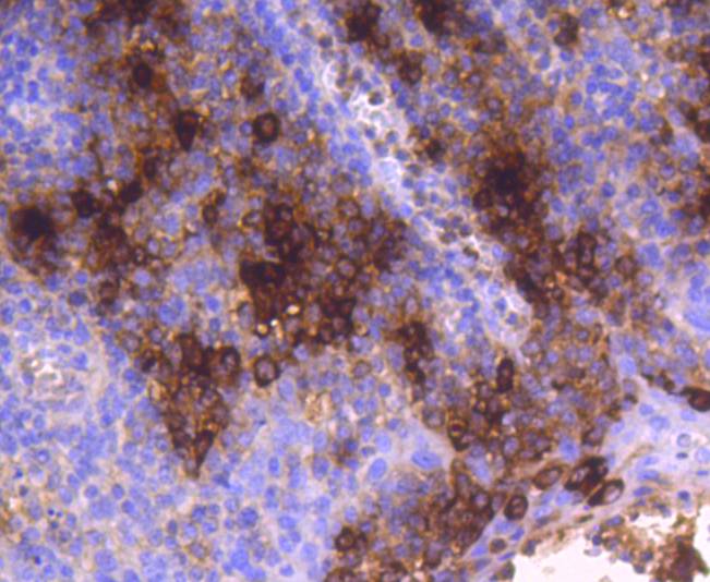
Immunohistochemical analysis of paraffin-embedded human tonsil tissue using anti-Mast Cell Tryptase antibody. Counter stained with hematoxylin.
Immunohistochemical analysis of paraffin-embedded human lung tissue using anti-Mast Cell Tryptase antibody. Counter stained with hematoxylin.
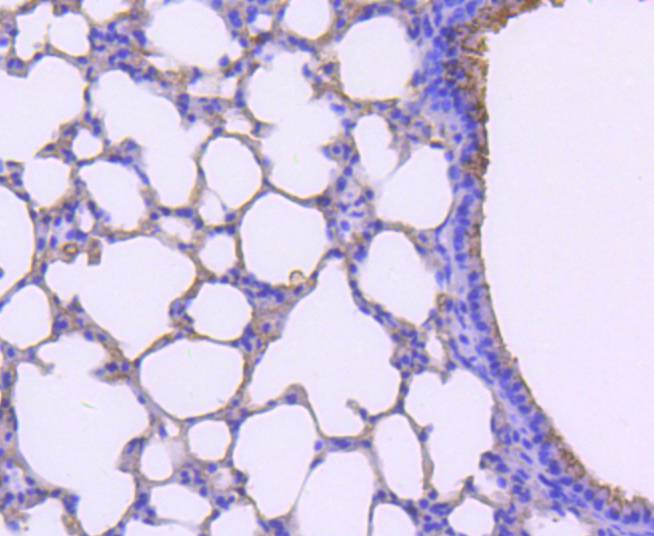
Immunohistochemical analysis of paraffin-embedded mouse lung tissue using anti-Mast Cell Tryptase antibody. Counter stained with hematoxylin.
Mast cells are connective tissue cells derived from blood-forming tissues that line arterial walls and secrete substances, which mediate inflammatory and immune responses. Mast cell chymase, known as CMA1, is a major secreted serine protease that is involved in vasoactive peptide generation, extracellular matrix degradation and regulation of gland secretion. The human chymase gene, which maps to human chromosome 14q11.2, encodes a preproenzyme with a 19-amino acid signal peptide, an acidic 2-amino acid propeptide and a 226-amino acid catalytic domain. Tryptases comprise a family of trypsin-like serine proteases that are enzymatically active as heparin-stabilized tetramers. There are four functional genes for tryptase: α I, β I, β II and γ I, which map to human chromosome 16p13.3, with β tryptases representing the main isoenzymes expressed in mast cells. Mast cell proteases are a family of rodent protein homologs to human tryptases that are specifically expressed in mast cells and may serve as highly specific markers in the analysis of mast cell heterogeneity, differentiation and function.
If you have published an article using product 48980, please notify us so that we can cite your literature.

 Yes
Yes