Product Detail
Product NameHLA-DR Rabbit mAb
Clone No.SR4525
Host SpeciesRecombinant Rabbit
ClonalityMonoclonal
PurificationProA affinity purified
ApplicationsWB, ICC, IHC, FC
Species ReactivityHuman;Mouse
Immunogen Descrecombinant protein
ConjugateUnconjugated
Other NamesDR alpha chain antibody DR alpha chain precursor antibody DRA_HUMAN antibody DRB1 antibody DRB4 antibody Histocompatibility antigen HLA DR alpha antibody HLA class II histocompatibility antigen antibody HLA class II histocompatibility antigen DR alpha chain antibody HLA DR1B antibody HLA DR3B antibody HLA DRA antibody HLA DRA1 antibody HLA DRB1 antibody HLA DRB3 antibody HLA DRB4 antibody HLA DRB5 antibody HLA-DRA antibody HLADR4B antibody HLADRA1 antibody HLADRB antibody Major histocompatibility complex class II DR alpha antibody Major histocompatibility complex class II DR beta 1 antibody Major histocompatibility complex class II DR beta 3 antibody Major histocompatibility complex class II DR beta 4 antibody Major histocompatibility complex class II DR beta 5 antibody MGC117330 antibody MHC cell surface glycoprotein antibody MHC class II antigen DRA antibody MHC II antibody MLRW antibody
Accession NoSwiss-Prot#:P01903
Uniprot
P01903
Gene ID
3122;
Calculated MW29 kDa
Formulation1*TBS (pH7.4), 1%BSA, 40%Glycerol. Preservative: 0.05% Sodium Azide.
StorageStore at -20˚C
Application Details
WB: 1:1,000-5,000
IHC: 1:50-1:200
ICC: 1:50-1:200
FC: 1:50-1:100
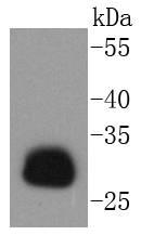
Western blot analysis of HLA-DR on Daudi cells lysates using anti-HLA-DR antibody at 1/1,000 dilution.
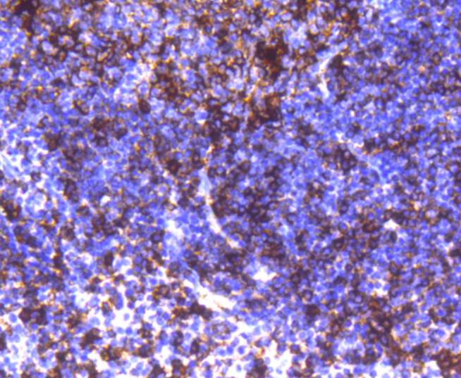
Immunohistochemical analysis of paraffin-embedded human tonsil tissue using anti-HLA-DR antibody. Counter stained with hematoxylin.
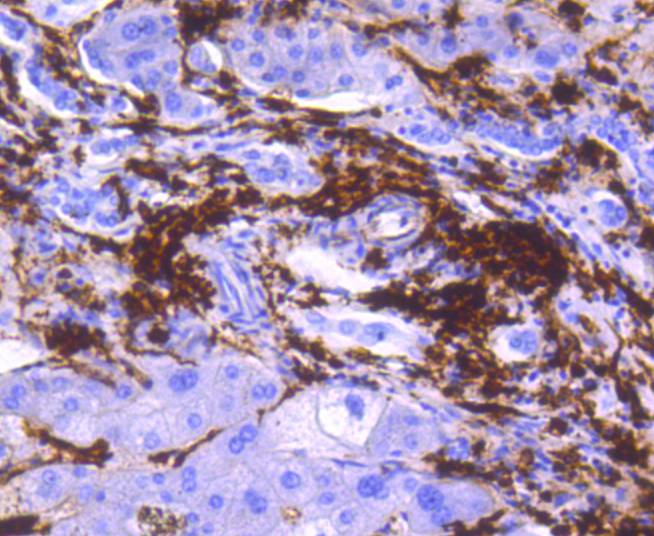
Immunohistochemical analysis of paraffin-embedded human liver tissue using anti-HLA-DR antibody. Counter stained with hematoxylin.
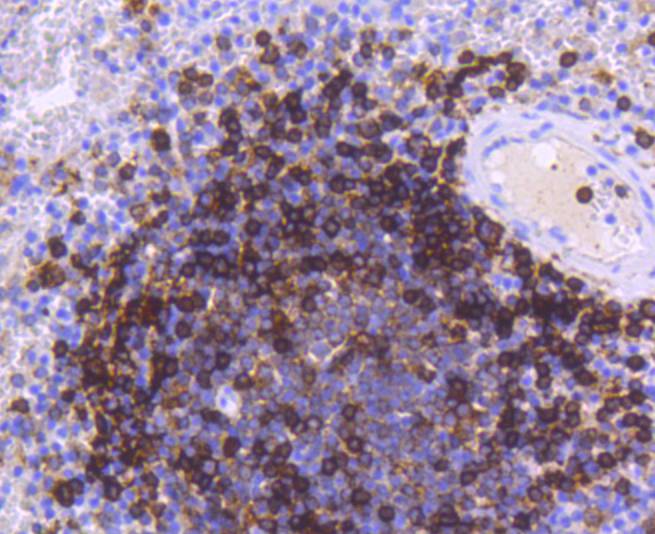
Immunohistochemical analysis of paraffin-embedded human spleen tissue using anti-HLA-DR antibody. Counter stained with hematoxylin.
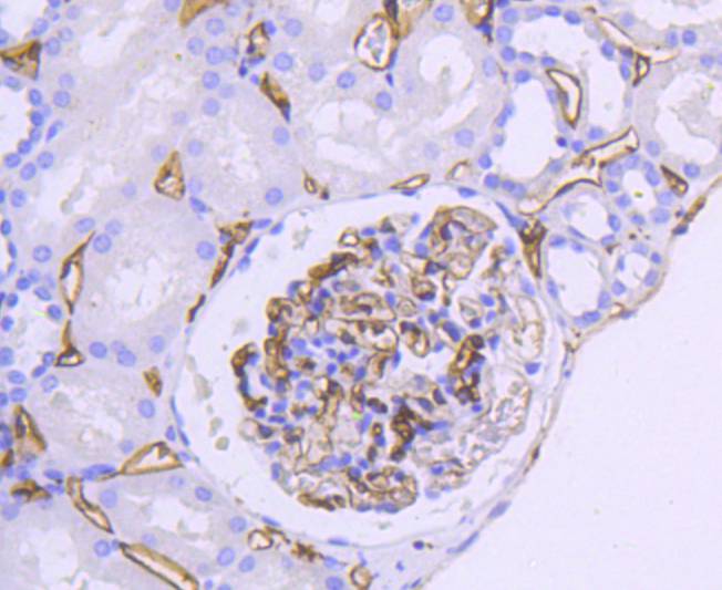
Immunohistochemical analysis of paraffin-embedded human kidney tissue using anti-HLA-DR antibody. Counter stained with hematoxylin.
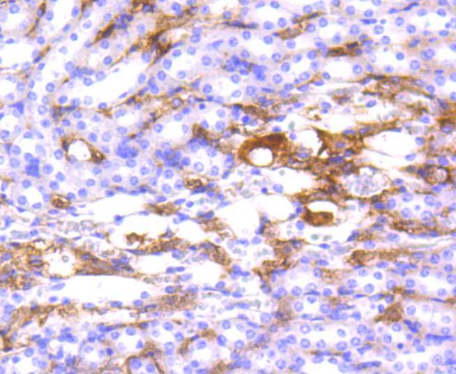
Immunohistochemical analysis of paraffin-embedded mouse kidney tissue using anti-HLA-DR antibody. Counter stained with hematoxylin.
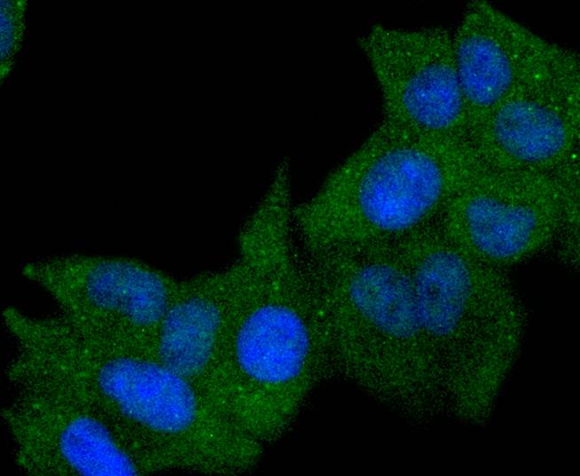
ICC staining HLA-DR in Hela cells (green). The nuclear counter stain is DAPI (blue). Cells were fixed in paraformaldehyde, permeabilised with 0.25% Triton X100/PBS.
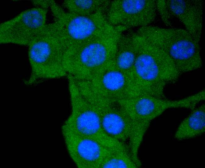
ICC staining HLA-DR in B16-F1 cells (green). The nuclear counter stain is DAPI (blue). Cells were fixed in paraformaldehyde, permeabilised with 0.25% Triton X100/PBS.
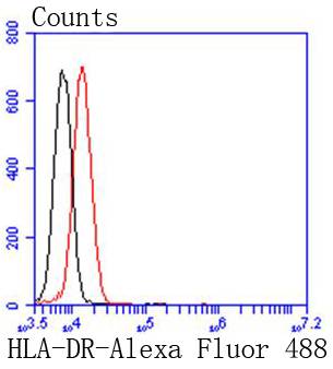
Flow cytometric analysis of Jurkat cells with HLA-DR antibody at 1/50 dilution (red) compared with an unlabelled control (cells without incubation with primary antibody; black). Alexa Fluor 488-conjugated goat anti rabbit IgG was used as the secondary antibody.
Major histocompatibility complex (MHC) class II molecules destined for presentation to CD4+ helper T cells is determined by two key events. These events include the dissociation of class II-associated invariant chain peptides (CLIP) from an antigen binding groove in MHC II α/β dimers through the activity of MHC molecules HLA-DM and -DO, and subsequent peptide antigen binding. Accumulating in endosomal/lysosomal compartments and on the surface of B cells, HLA-DM, -DO molecules regulate the dissociation of CLIP and the subsequent binding of exogenous peptides to HLA class II molecules (HLA-DR, -DQ and -DP) by sustaining a conformation that favors peptide exchange. RFLP analysis of HLA-DM genes from rheumatoid arthritis (RA) patients suggests that certain polymorphisms are genetic factors for RA susceptibility. HLA-B belongs to the HLA class I heavy chain paralogs. Class I molecules play a central role in the immune system by presenting peptides derived from the endoplasmic reticulum lumen. HLA-B and -C can form heterodimers consisting of a membrane anchored heavy chain and a light chain (β-2-Microglobulin). Polymorphisms yield hundreds of HLA-B and -C alleles.
If you have published an article using product 48982, please notify us so that we can cite your literature.


 Yes
Yes


