Product Detail
Product NameATG9A Rabbit mAb
Clone No.SC67-05
Host SpeciesRecombinant Rabbit
Clonality Monoclonal
PurificationProA affinity purified
ApplicationsWB, ICC, IHC, IP
Species ReactivityHu, Ms, Rt
Immunogen Descrecombinant protein
ConjugateUnconjugated
Other NamesAPG9 autophagy 9-like 1 antibody APG9 like 1 antibody APG9-like 1 antibody APG9L1 antibody ATG9 antibody ATG9 autophagy related 9 homolog A antibody ATG9 autophagy related 9 homolog A (S. cerevisiae) antibody ATG9A antibody ATG9A_HUMAN antibody Autophagy 9-like 1 protein antibody Autophagy related protein 9A antibody Autophagy-related protein 9A antibody mATG9 antibody MGD3208 antibody OTTHUMP00000206046 antibody OTTHUMP00000206048 antibody OTTHUMP00000206049 antibody OTTHUMP00000206062 antibody
Accession NoSwiss-Prot#:Q7Z3C6
Uniprot
Q7Z3C6
Gene ID
79065;
Calculated MW94 kDa
Formulation1*TBS (pH7.4), 1%BSA, 40%Glycerol. Preservative: 0.05% Sodium Azide.
StorageStore at -20˚C
Application Details
WB: 1:1,000
IHC: 1:50-1:200
ICC: 1:50-1:200
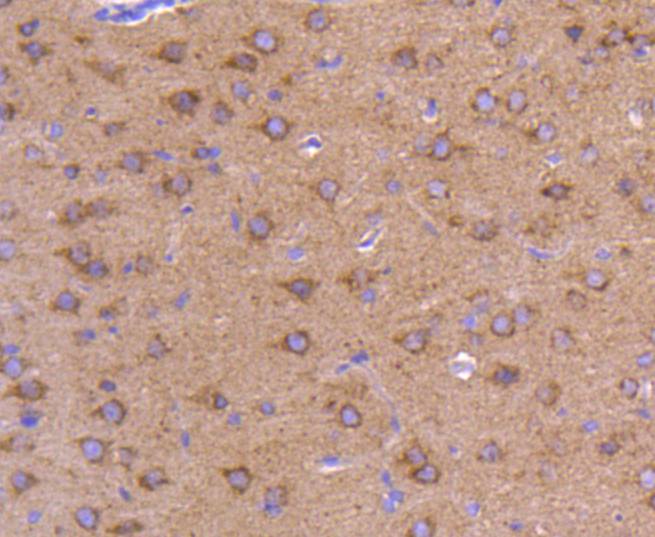
Immunohistochemical analysis of paraffin-embedded rat brain tissue using anti-ATG9A antibody. Counter stained with hematoxylin.
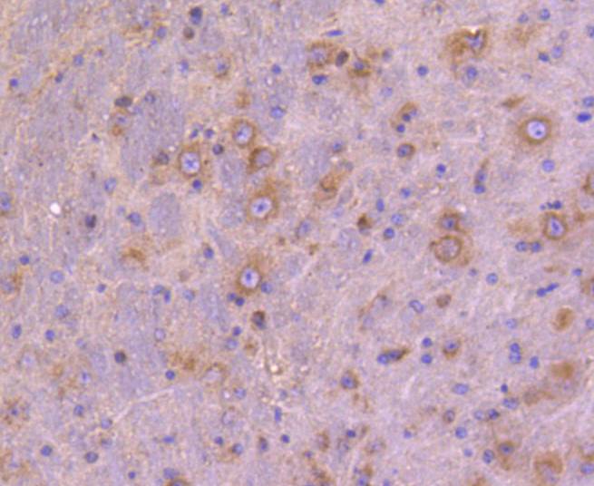
Immunohistochemical analysis of paraffin-embedded mouse brain tissue using anti-ATG9A antibody. Counter stained with hematoxylin.
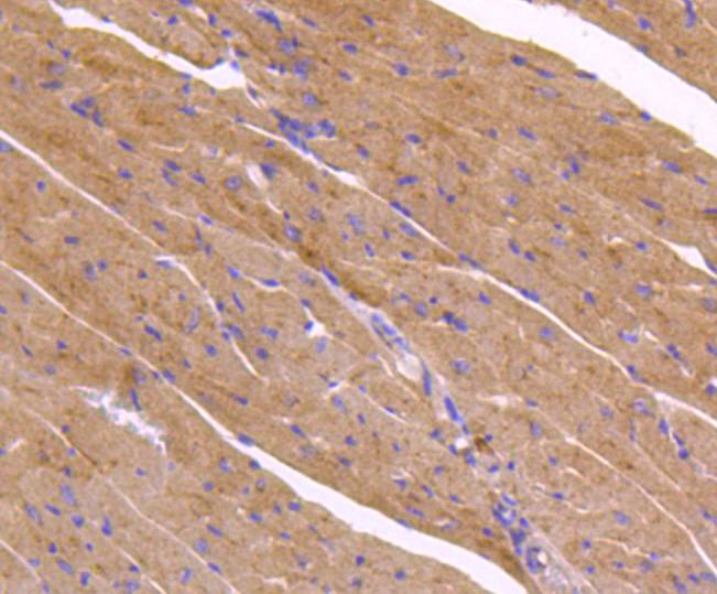
Immunohistochemical analysis of paraffin-embedded mouse heart tissue using anti-ATG9A antibody. Counter stained with hematoxylin.
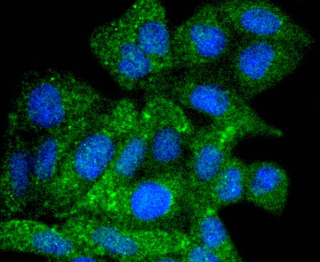
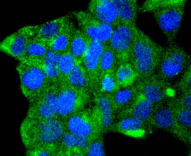
ICC staining ATG9A in Hela cells (green). The nuclear counter stain is DAPI (blue). Cells were fixed in paraformaldehyde, permeabilised with 0.25% Triton X100/PBS.
ICC staining ATG9A in 293 cells (green). The nuclear counter stain is DAPI (blue). Cells were fixed in paraformaldehyde, permeabilised with 0.25% Triton X100/PBS.
Autophagy, a process that results in the lysosomal-dependent degradation of cytosolic compartments, is carried out by the autophagosome, which is a double-membrane vesicle whose formation is catalyzed by several autophagy-related gene (Atg) proteins. Atg9a (autophagy-related protein 9A), also known as APG9-like 1, is a 839 amino acid, multi-pass membrane protein that localizes to the pre-autophagosomal structure (PAS). Isolation membranes are suggested to originate from the PAS, enwrapping cytoplasmic components to form a double membrane autophagosome, which then fuses with the vacuole. Ubiquitously expressed in human adult tissues, Atg9a cycles between the Golgi and endosomes and, with the autophagosome-specific marker LC3, plays a critical role in starvation-induced autophagosome formation. Three isoforms of Atg9a exist as a result of alternative splicing events.
If you have published an article using product 48988, please notify us so that we can cite your literature.


 Yes
Yes


