Welcome to Sabbiotech!     
Email:info@signalwayantibody.com
Location: Home > Recombinant Rabbit Monoclonal Antibodies > TCF7L2 Rabbit mAb

TCF7L2 Rabbit mAb#48990

Availability: Yes
Total Amount:$
Product Detail

Product NameTCF7L2 Rabbit mAb

Clone No.SC06-90

Host SpeciesRecombinant Rabbit

Clonality Monoclonal

PurificationProA affinity purified

ApplicationsWB, ICC/IF, IHC, IP, FC

Species ReactivityHu, Ms, Rt

Immunogen Descrecombinant protein

ConjugateUnconjugated

Other NamesHMG box transcription factor 4 antibody
hTCF 4 antibody
hTCF-4 antibody
T cell factor 4 antibody
T cell specific HMG box antibody
T cell specific transcription factor 4 antibody
T-cell factor 4 antibody
T-cell-specific transcription factor 4 antibody
TCF 4 antibody
TCF-4 antibody
TCF4 antibody
TCF7L2 antibody
TCF7L2 protein antibody
TF7L2_HUMAN antibody
Transcription factor 7 like 2 antibody
Transcription factor 7 like 2 T cell specific HMG box antibody
Transcription factor 7-like 2 antibody

Accession NoSwiss-Prot#:Q9NQB0

Uniprot Q9NQB0

Gene ID 6934;

Calculated MW68 kDa

Formulation1*TBS (pH7.4), 1%BSA, 40%Glycerol. Preservative: 0.05% Sodium Azide.

StorageStore at -20˚C

Application Details
WB: 1:500-1:1000
IHC: 1:50-1:200
ICC: 1:50-1:200

FC: 1:50-1:100
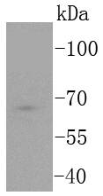
Western blot analysis of TCF7L2 on Jurkat cells lysates using anti-TCF7L2 antibody at 1/1,000 dilution.
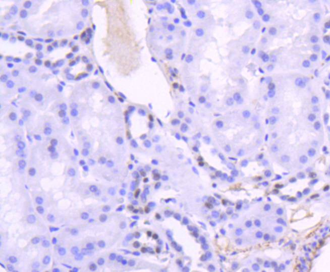
Immunohistochemical analysis of paraffin-embedded human kidney tissue using anti-TCF7L2 antibody. Counter stained with hematoxylin.
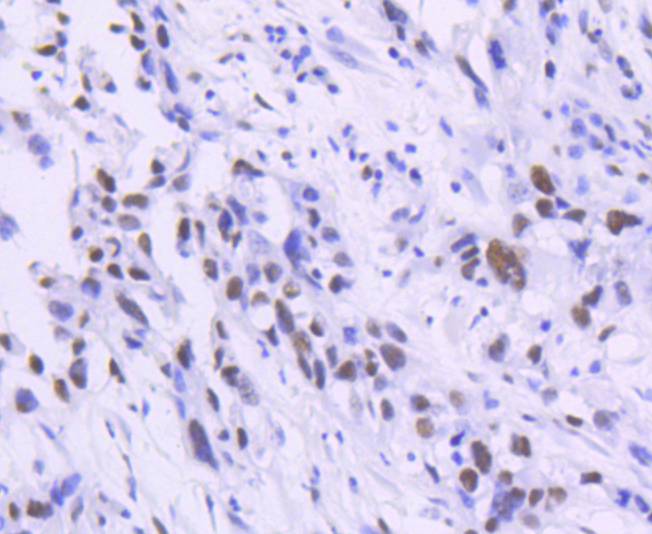
Immunohistochemical analysis of paraffin-embedded human breast carcinoma tissue using anti-TCF7L2 antibody. Counter stained with hematoxylin.
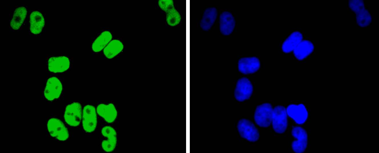
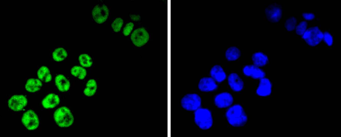
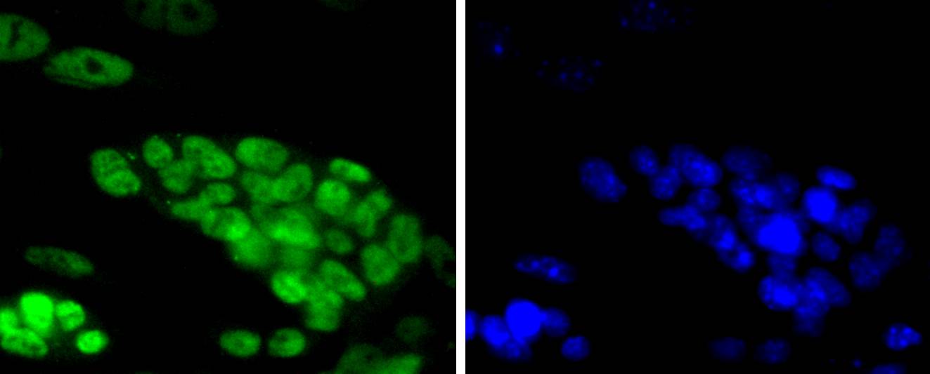
ICC staining TCF7L2 in Hela cells (green). The nuclear counter stain is DAPI (blue). Cells were fixed in paraformaldehyde, permeabilised with 0.25% Triton X100/PBS.
ICC staining TCF7L2 in SW480 cells (green). The nuclear counter stain is DAPI (blue). Cells were fixed in paraformaldehyde, permeabilised with 0.25% Triton X100/PBS.
ICC staining TCF7L2 in D3 cells (green). The nuclear counter stain is DAPI (blue). Cells were fixed in paraformaldehyde, permeabilised with 0.25% Triton X100/PBS.
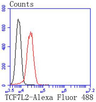
Flow cytometric analysis of Jurkat cells with TCF7L2 antibody at 1/50 dilution (red) compared with an unlabelled control (cells without incubation with primary antibody; black). Alexa Fluor 488-conjugated goat anti rabbit IgG was used as the secondary antibody.
T cell factors (TCFs) comprise a family of DNA-binding transcriptional activators that are essential for lymphoid cell development. These transcription factors are activated by the Wnt-1 and Wingless pathways and are characterized by the presence of a conserved protein motif, the high mobility group (HMG) 1 box, which mediates DNA binding. TCF-4 mainly localizes in the cytoplasm and is transported into the nucleus directly bound to β-catenin in a cooperative manner. This TCF-4/β-catenin complex induces expression of Wnt target genes, including multiple cancer-associated genes. c-Jun also interacts with TCF-4 and β-catenin, and the phosphorylation-dependent interaction between c-Jun and TCF4 regulates intestinal tumorigenesis by integrating JNK and APC/β-catenin. TCF-4 is also implicated in bipolar affective disorder.

If you have published an article using product 48990, please notify us so that we can cite your literature.

NOTE

Application

  • WBWestern Blotting
  • IHCImmunohistochemistry
  • IFImmunofluorescence
  • ICCImmunocytochemistry
  • FCFlow Cytometry
  • IPImmunoprecipitation
  • EELISA
  • DBDot Blotting
  • ChIPChromatin Immunoprecipitation
  • GICAGold Immunochromatography Assay
  • NCNegative Control

Species Reactivity

  • HuHuman
  • MsMouse
  • RtRat
  • DmDrosophila melanogaster
  • CCaenorhabditis elegans
  • MkMonkey
  • RbRabbit
  • BBovine
  • DDog
  • PPig
  • HmHamster
  • ChHmChinese Hamster
  • ChkChicken
  • ShpSheep
;