Welcome to Sabbiotech!     
Email:info@signalwayantibody.com
Location: Home > Recombinant Rabbit Monoclonal Antibodies > GAP43 Rabbit mAb

GAP43 Rabbit mAb#49013

Availability: Yes
Total Amount:$
Product Detail

Product NameGAP43 Rabbit mAb

Clone No.SC60-06

Host SpeciesRecombinant Rabbit

Clonality Monoclonal

PurificationProA affinity purified

ApplicationsWB, ICC/IF, IHC, IP, FC

Species ReactivityHu, Ms, Rt

Immunogen Descrecombinant protein

ConjugateUnconjugated

Other NamesAxonal membrane protein GAP 43 antibody
Axonal membrane protein GAP-43 antibody
B 50 antibody
Calmodulin binding protein P 57 antibody
F1 antibody
GAP 43 antibody
GAP43 antibody
Growth Associated Protein 43 antibody
Growth-associated protein 43 antibody
Nerve Growth Related Peptide antibody
Nerve growth related peptide GAP43 antibody
NEUM_HUMAN antibody
Neural phosphoprotein B 50 antibody
Neural phosphoprotein B-50 antibody
Neuromodulin antibody
Neuron growth associated protein 43 antibody
PP46 antibody
Protein F1 antibody
QtrA-11580 antibody
QtrA-13071 antibody

Accession NoSwiss-Prot#:P17677

Uniprot P17677

Gene ID 2596;

Calculated MW48 kDa

Formulation1*TBS (pH7.4), 1%BSA, 40%Glycerol. Preservative: 0.05% Sodium Azide.

StorageStore at -20˚C

Application Details
WB: 1:2,000-1:5,000
IHC: 1:100-1:500
ICC: 1:100-1:500

FC: 1:50-1:100
Western blot analysis of GAP43 on mouse brain lysates using anti-GAP43 antibody at 1/1,000 dilution.
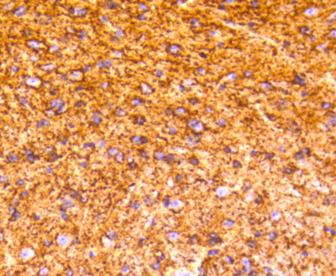
Immunohistochemical analysis of paraffin-embedded mouse brain tissue using anti-GAP43 antibody. Counter stained with hematoxylin.
ICC staining GAP43 in Hela cells (green). The nuclear counter stain is DAPI (blue). Cells were fixed in paraformaldehyde, permeabilised with 0.25% Triton X100/PBS.
ICC staining GAP43 in SHG-44 cells (green). The nuclear counter stain is DAPI (blue). Cells were fixed in paraformaldehyde, permeabilised with 0.25% Triton X100/PBS.
Flow cytometric analysis of SH-SY-5y cells with GAP43 antibody at 1/50 dilution (red) compared with an unlabelled control (cells without incubation with primary antibody; black). Alexa Fluor 488-conjugated goat anti rabbit IgG was used as the secondary antibody.
GAP-43 (growth associated protein 43, B-50, PP46, calmodulin-binding protein P-57, neuromodulin, neuron growth-associated protein 43, protein F1 is a crucial component for regenerative response in the nervous system. It is present at high levels in neuronal growth cones during development and axonal regeneration. GAP-43 is normally produced by neurons during developmental growth and axonal regeneration, but it is also expressed in specific regions of the normal adult nervous system. The neuron-specific Elav/Hu family member, HuD, interacts with and stabilizes GAP-43 mRNA in developing neurons and leads to increased levels of GAP-43 protein. Heterozygous GAP-43 knockout mice with GAP-43 levels reduced by one-half dipslay significant memory impairments in cued conditioning or on tests of nociceptive or auditory perception.

If you have published an article using product 49013, please notify us so that we can cite your literature.

et al,Maf1 is an intrinsic suppressor against spontaneous neural repair and functional recovery after ischemic stroke. In J Adv Res on 2022 Nov 17 by Chi Kwan Tsang, Qiongjie Mi,et al..PMID:36402285 , (2022),
PMID: 36402285
et al,Maf1 is an intrinsic suppressor against spontaneous neural repair and functional recovery after ischemic strokeInJ Adv ResOn2023 SepbyChi Kwan Tsang , Qiongjie Mi et al..PMID:36402285 , (2023),
PMID: 36402285

NOTE

Application

  • WBWestern Blotting
  • IHCImmunohistochemistry
  • IFImmunofluorescence
  • ICCImmunocytochemistry
  • FCFlow Cytometry
  • IPImmunoprecipitation
  • EELISA
  • DBDot Blotting
  • ChIPChromatin Immunoprecipitation
  • GICAGold Immunochromatography Assay
  • NCNegative Control

Species Reactivity

  • HuHuman
  • MsMouse
  • RtRat
  • DmDrosophila melanogaster
  • CCaenorhabditis elegans
  • MkMonkey
  • RbRabbit
  • BBovine
  • DDog
  • PPig
  • HmHamster
  • ChHmChinese Hamster
  • ChkChicken
  • ShpSheep
;