Product Detail
Product NameNSE Rabbit mAb
Clone No.SC06-28
Host SpeciesRecombinant Rabbit
Clonality Monoclonal
PurificationProA affinity purified
ApplicationsWB, ICC, IHC
Species ReactivityHu, Ms, Rt, zebrafish
Immunogen Descrecombinant protein
ConjugateUnconjugated
Other Names2 phospho D glycerate hydrolyase antibody
2-phospho-D-glycerate hydro-lyase antibody
Eno 2 antibody
ENO2 antibody
ENOG antibody
ENOG_HUMAN antibody
Enolase 2 (gamma, neuronal) antibody
Enolase 2 antibody
Enolase 2 gamma neuronal antibody
Enolase2 antibody
Epididymis secretory protein Li 279 antibody
Gamma enolase antibody
Gamma-enolase antibody
HEL S 279 antibody
Neural enolase antibody
Neuron specific enolase antibody
Neuron specific gamma enolase antibody
Neuron-specific enolase antibody
Neurone specific enolase antibody
NSE antibody
Accession NoSwiss-Prot#:P09104
Uniprot
P09104
Gene ID
2026;
Calculated MW47 kDa
Formulation1*TBS (pH7.4), 1%BSA, 40%Glycerol. Preservative: 0.05% Sodium Azide.
StorageStore at -20˚C
Application Details
WB: 1:1,000-1:2,000
IHC: 1:50-1:200
ICC: 1:50-1:200
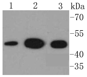
Western blot analysis of NSE on different lysates using anti-NSE antibody at 1/1,000 dilution. Positive control:
Lane 1: HepG2
Lane 2: Hela
Lane 3: 293
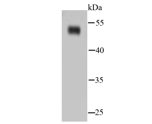
Western blot analysis of NSE on hybrid fish (crucian-carp) brain tissue lysates using anti-NSE antibody at 1/500 dilution.
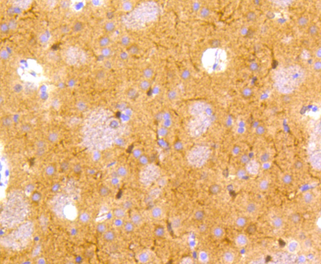
Immunohistochemical analysis of paraffin-embedded rat brain tissue using anti-NSE antibody. Counter stained with hematoxylin.
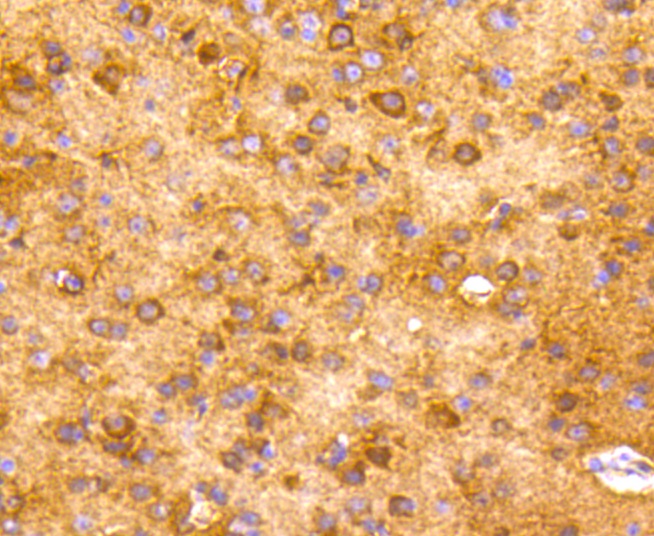
Immunohistochemical analysis of paraffin-embedded human tonsil tissue using anti-NSE antibody. Counter stained with hematoxylin.
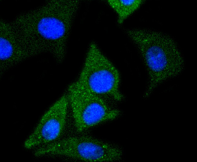
ICC staining NSE in SH-SY-5Y cells (green). The nuclear counter stain is DAPI (blue). Cells were fixed in paraformaldehyde, permeabilised with 0.25% Triton X100/PBS.
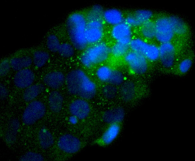
ICC staining NSE in 293 cells (green). The nuclear counter stain is DAPI (blue). Cells were fixed in paraformaldehyde, permeabilised with 0.25% Triton X100/PBS.
Enolases have been characterized as highly conserved cytoplasmic glycolytic enzymes that may be involved in differentiation. Three isoenzymes have been identified, α Enolase, β Enolase and γ Enolase. α Enolase expression has been detected on most tissues, whereas β Enolase is expressed predominantly in muscle tissue and γ Enolase is detected only in nervous tissue. These isoforms exist as both homodimers and heterodimers, and they play a role in converting phosphoglyceric acid to phosphenolpyruvic acid in the glycolytic pathway.
If you have published an article using product 49015, please notify us so that we can cite your literature.


 Yes
Yes


