Product Detail
Product NameTNFAIP3 Rabbit mAb
Clone No.SN07-31
Host SpeciesRecombinant Rabbit
Clonality Monoclonal
PurificationProA affinity purified
ApplicationsWB, ICC/IF, IHC, FC
Species ReactivityHu
Immunogen Descrecombinant protein
ConjugateUnconjugated
Other NamesA20 antibody AISBL antibody MGC104522 antibody MGC138687 antibody MGC138688 antibody OTU domain containing protein 7C antibody OTU domain-containing protein 7C antibody OTUD7C antibody Putative DNA binding protein A20 antibody Putative DNA-binding protein A20 antibody TNAP3_HUMAN antibody TNF alpha-induced protein 3 antibody TNFA1P2 antibody TNFAIP 3 antibody TNFAIP3 (A20) antibody TNFAIP3 antibody Tumor necrosis factor alpha induced protein 3 antibody Tumor necrosis factor alpha-induced protein 3 antibody Tumor necrosis factor induced protein 3 antibody Tumor necrosis factor inducible protein A20 antibody tumor necrosis factor, alpha-induced protein 3 antibody Zinc finger protein A20 antibody
Accession NoSwiss-Prot#:P21580
Uniprot
P21580
Gene ID
7128;
Calculated MW90 kDa
Formulation1*TBS (pH7.4), 1%BSA, 40%Glycerol. Preservative: 0.05% Sodium Azide.
StorageStore at -20˚C
Application Details
WB: 1:1,000
IHC: 1:50-1:200
ICC: 1:100-1:500
FC: 1:50-1:100
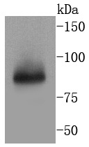
Western blot analysis of TNFAIP3 on Jurkat cells lysates using anti-TNFAIP3 antibody at 1/1,000 dilution.
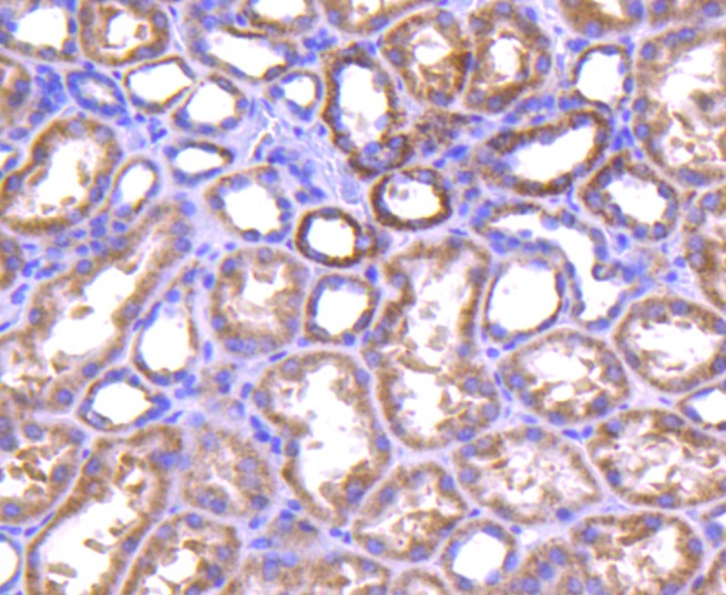
Immunohistochemical analysis of paraffin-embedded human kidney tissue using anti-TNFAIP3 antibody. Counter stained with hematoxylin.
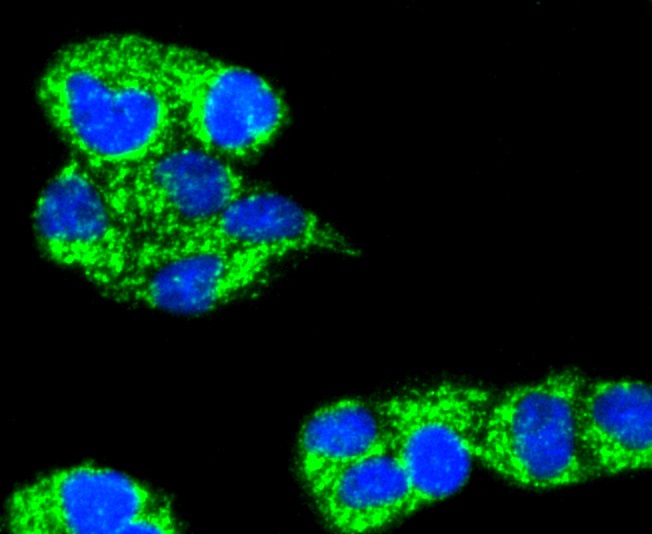
ICC staining TNFAIP3 in Hela cells (green). The nuclear counter stain is DAPI (blue). Cells were fixed in paraformaldehyde, permeabilised with 0.25% Triton X100/PBS.
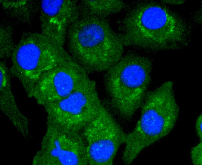
ICC staining TNFAIP3 in A549 cells (green). The nuclear counter stain is DAPI (blue). Cells were fixed in paraformaldehyde, permeabilised with 0.25% Triton X100/PBS.
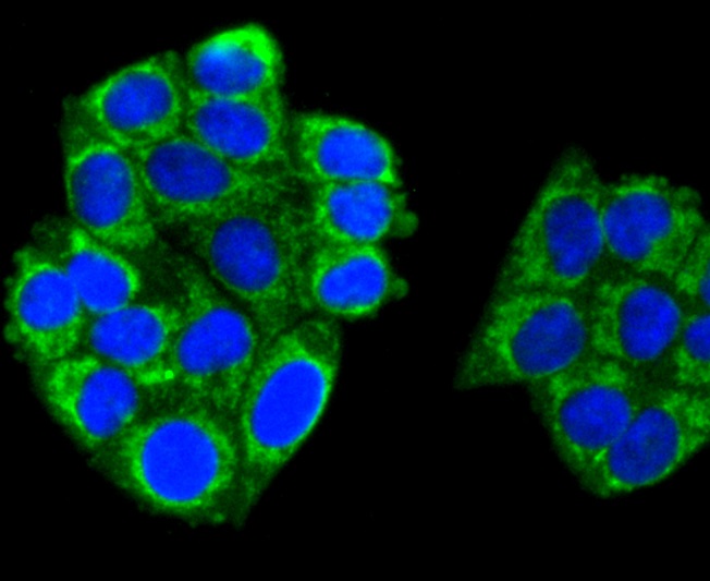
ICC staining TNFAIP3 in HepG2 cells (green). The nuclear counter stain is DAPI (blue). Cells were fixed in paraformaldehyde, permeabilised with 0.25% Triton X100/PBS.
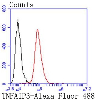
Flow cytometric analysis of HepG2 cells with TNFAIP3 antibody at 1/50 dilution (red) compared with an unlabelled control (cells without incubation with primary antibody; black). Alexa Fluor 488-conjugated goat anti rabbit IgG was used as the secondary antibody
A20 is a Cys2/Cys2 zinc finger protein that is induced by a variety of inflammatory stimuli and regulates gene expression. Specifically, A20 is induced by tumor necrosis factor (TNF) and interleukin 1 (IL-1), and acts as a negative regulator of nuclear factor κ B (NFκB) gene expression. By inhibiting NFκB activation, A20 plays a critical role in terminating NFκB responses to various stimuli. Although the C-terminal region of A20 contains seven zinc finger domains, only four of these domains are required for in vitro inhibition of TNF-induced NFκB activation. A20 also interacts with several other proteins, such as TRAF2, TRAF6 and IκB kinase (IKK) γ protein, and can thereby inhibit cell death. TXBP151, a novel A20-binding protein, may mediate the anti-apoptotic activity of A20. Involved in the negative feedback regulation of signal transduction, A20 and A20-binding proteins may be useful as novel therapeutic tools in the treatment of a variety of diseases.
If you have published an article using product 49053, please notify us so that we can cite your literature.


 Yes
Yes


