Product Detail
Product NameCytokeratin 13 Rabbit mAb
Clone No.SN71-09
Host SpeciesRecombinant Rabbit
Clonality Monoclonal
PurificationProA affinity purified
ApplicationsWB, ICC, IHC
Species ReactivityHu, Ms, zebrafish
Immunogen Descrecombinant protein
ConjugateUnconjugated
Other Names47 kDa cytokeratin antibody
CK-13 antibody
CK13 antibody
Cytokeratin 13 antibody
Cytokeratin-13 antibody
K13 antibody
K1C13_HUMAN antibody
Ka13 antibody
Keratin 13 antibody
Keratin antibody
keratin type I cytoskeletal 13 antibody
Keratin-13 antibody
Krt-1.13 antibody
Krt1-13 antibody
KRT13 antibody
MGC161462 antibody
MGC3781 antibody
type I cytoskeletal 13 antibody
Type I keratin Ka13 antibody
WSN2 antibody
Accession NoSwiss-Prot#:P13646
Uniprot
P13646
Gene ID
3860;
Calculated MW50 kDa
Formulation1*TBS (pH7.4), 1%BSA, 40%Glycerol. Preservative: 0.05% Sodium Azide.
StorageStore at -20˚C
Application Details
WB: 1:1,000-5,000
IHC: 1:50-1:200
ICC: 1:50-1:200
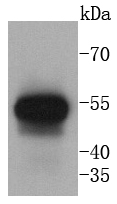
Western blot analysis of Cytokeratin 13 on human lung lysates using anti-Cytokeratin 13 antibody at 1/1,000 dilution.
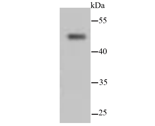
Western blot analysis of Cytokeratin 13 hybrid fish (crucian-carp) brain tissue lysate using anti-Cytokeratin 13 antibody at 1/500 dilution.
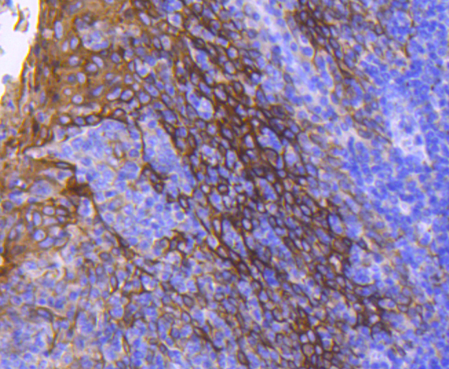
Immunohistochemical analysis of paraffin-embedded human tonsil tissue using anti-Cytokeratin 13 antibody. Counter stained with hematoxylin.
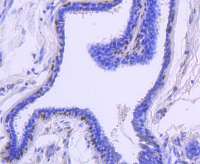
Immunohistochemical analysis of paraffin-embedded human breast carcinoma tissue using anti-Cytokeratin 13 antibody. Counter stained with hematoxylin.
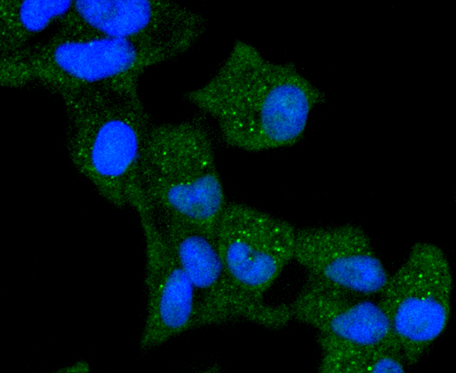
ICC staining Cytokeratin 13 in Hela cells (green). The nuclear counter stain is DAPI (blue). Cells were fixed in paraformaldehyde, permeabilised with 0.25% Triton X100/PBS.
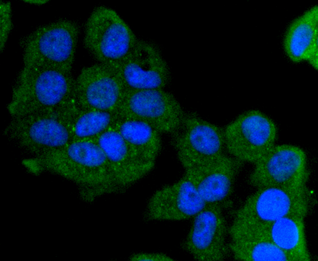
ICC staining Cytokeratin 13 in MCF-7 cells (green). The nuclear counter stain is DAPI (blue). Cells were fixed in paraformaldehyde, permeabilised with 0.25% Triton X100/PBS.
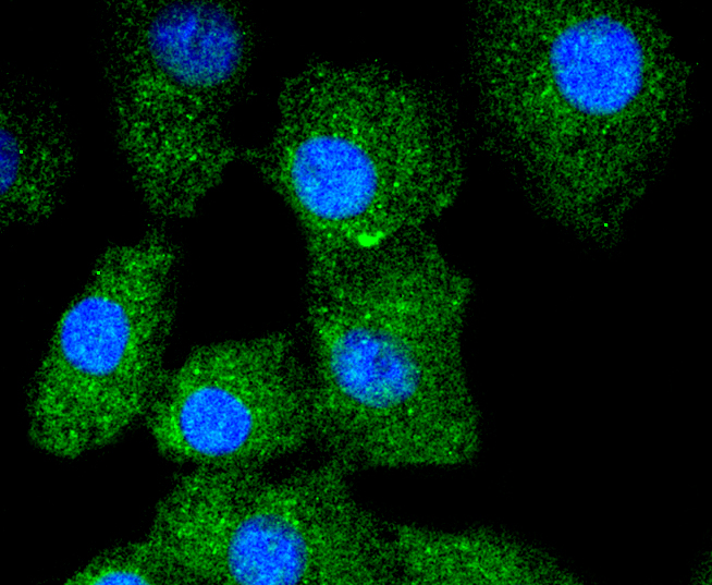
ICC staining Cytokeratin 13 in A549 cells (green). The nuclear counter stain is DAPI (blue). Cells were fixed in paraformaldehyde, permeabilised with 0.25% Triton X100/PBS.
Cytokeratins comprise a diverse group of intermediate filament proteins (IFPs) that are expressed as pairs in both keratinized and non-keratinized epithelial tissue. Cytokeratins play a critical role in differentiation and tissue specialization and function to maintain the overall structural integrity of epithelial cells. Cytokeratins have been found to be useful markers of tissue differentiation, which is directly applicable to the characterization of malignant tumors. Cytokeratins 10 and 13 are present in the cytoskeletal region of a subset of squamous cell carcinomas. Cytokeratin 13 belongs to the intermediate filament family and is a heterotetramer of two type I acidic and two type II basic keratins. It is generally associated with cytokeratin 4. Defects in the KRT13 gene are a cause of white sponge nevus of cannon (WSN), a rare autosomal dominant disorder which predominantly affects noncornified stratified squamous epithelia and is characterized by the presence of soft, white and spongy plaques in the oral mucosa.
If you have published an article using product 49069, please notify us so that we can cite your literature.


 Yes
Yes


