Product Detail
Product NameCD13 Rabbit mAb
Clone No.SN71-04
Host SpeciesRecombinant Rabbit
Clonality Monoclonal
PurificationProA affinity purified
ApplicationsWB, ICC, IHC, IP
Species ReactivityHu, Ms, Rt
Immunogen Descrecombinant protein
ConjugateUnconjugated
Other NamesAlanyl (membrane) aminopeptidase antibody Alanyl aminopeptidase antibody Aminopeptidase M antibody Aminopeptidase N antibody AMPN_HUMAN antibody ANPEP antibody AP M antibody AP N antibody AP-M antibody AP-N antibody APN antibody CD 13 antibody CD13 antibody CD13 antigen antibody gp150 antibody hAPN antibody LAP 1 antibody LAP1 antibody Microsomal aminopeptidase antibody Myeloid plasma membrane glycoprotein CD13 antibody p150 antibody PEPN antibody
Accession NoSwiss-Prot#:P15144
Uniprot
P15144
Gene ID
290;
Calculated MW150 kDa
Formulation1*TBS (pH7.4), 1%BSA, 40%Glycerol. Preservative: 0.05% Sodium Azide.
StorageStore at -20˚C
Application Details
WB: 1:1,000-5,000
IHC: 1:100-1:500
ICC: 1:100-1:500
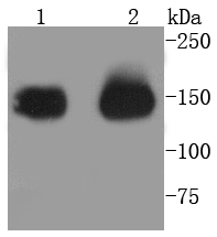
Western blot analysis of CD13 on different lysates using anti-CD13 antibody at 1/1,000 dilution. Positive control: Lane 1: THP-1 Lane 2: Human kidney
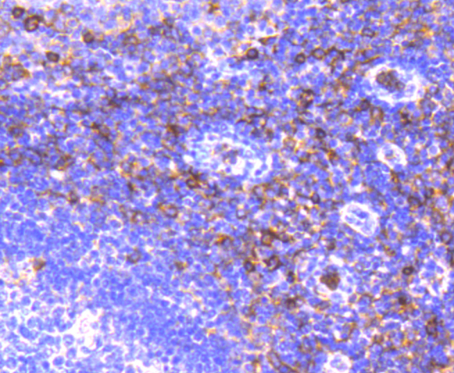
Immunohistochemical analysis of paraffin-embedded human tonsil tissue using anti-CD13 antibody. Counter stained with hematoxylin.
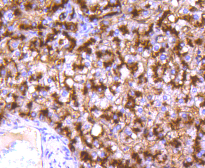
Immunohistochemical analysis of paraffin-embedded human liver tissue using anti-CD13 antibody. Counter stained with hematoxylin.
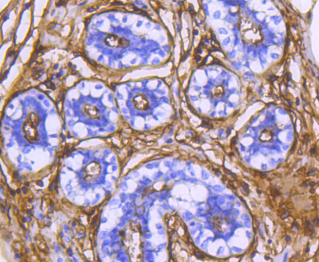
Immunohistochemical analysis of paraffin-embedded human breast tissue using anti-CD13 antibody. Counter stained with hematoxylin.
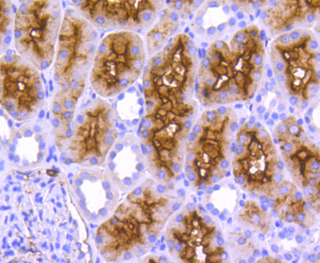
Immunohistochemical analysis of paraffin-embedded human kidney tissue using anti-CD13 antibody. Counter stained with hematoxylin.
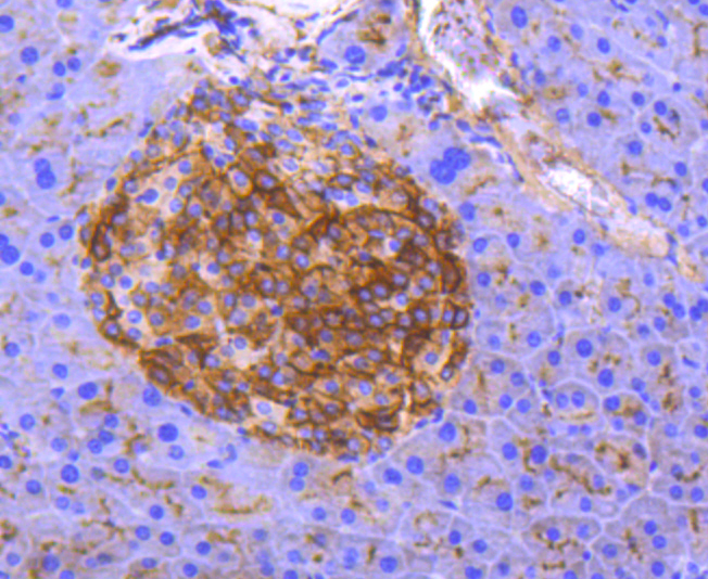
Immunohistochemical analysis of paraffin-embedded mouse pancreas tissue using anti-CD13 antibody. Counter stained with hematoxylin.
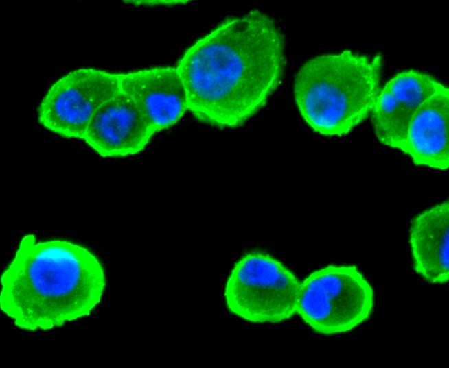
ICC staining CD13 in Hela cells (green). The nuclear counter stain is DAPI (blue). Cells were fixed in paraformaldehyde, permeabilised with 0.25% Triton X100/PBS
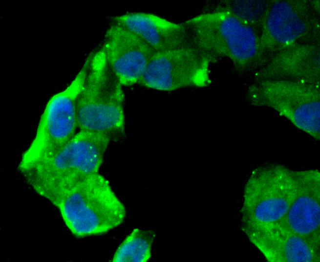
ICC staining CD13 in HepG2 cells (green). The nuclear counter stain is DAPI (blue). Cells were fixed in paraformaldehyde, permeabilised with 0.25% Triton X100/PBS.
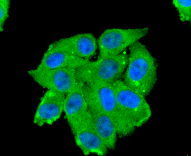
ICC staining CD13 in PANC-1 cells (green). The nuclear counter stain is DAPI (blue). Cells were fixed in paraformaldehyde, permeabilised with 0.25% Triton X100/PBS.
CD13, or aminopeptidase N, is a type II transmembrane glycoprotein that is expressed on most cells of Myeloid origin, including monocytes, basophils, eosinophils, neutrophils and Myeloid leukemias. CD13 is also found on certain epithelial cells, fibroblasts and osteoclasts. CD13 acts as a zinc-binding metalloprotease that plays a role in digestion and may function in the inactivation of some regulatory peptides such as enkephalins. CD13 may play a role in the invasion of cancer cells by enhancing their invasive capacity and metastatic behavior. The activity of CD13 can be inactivated using specific inhibitors that evoke apoptosis of CD13-positive cancer cells. Basic fibroblast growth factor (bFGF) expression upregulates CD13 expression in human melanoma cells by activating both the Myeloid and the epithelial CD13 promoter.
If you have published an article using product 49076, please notify us so that we can cite your literature.


 Yes
Yes


