Product Detail
Product NameTMS1 Rabbit mAb
Clone No.SN07-10
Host SpeciesRecombinant Rabbit
Clonality Monoclonal
PurificationProA affinity purified
ApplicationsWB, ICC/IF, FC
Species ReactivityHu
Immunogen Descrecombinant protein
ConjugateUnconjugated
Other NamesApoptosis associated speck like protein containing a CARD antibody Apoptosis-associated speck-like protein containing a CARD antibody ASC antibody ASC_HUMAN antibody CARD 5 antibody CARD5 antibody Caspase recruitment domain containing protein 5 antibody Caspase recruitment domain protein 5 antibody Caspase recruitment domain-containing protein 5 antibody hASC antibody MGC10332 antibody PYCARD antibody PYD and CARD domain containing antibody PYD and CARD domain containing protein antibody PYD and CARD domain-containing protein antibody Target of methylation induced silencing 1 antibody Target of methylation-induced silencing 1 antibody TMS 1 antibody TMS antibody TMS1 antibody
Accession NoSwiss-Prot#:Q9ULZ3
Uniprot
Q9ULZ3
Gene ID
29108;
Calculated MW22 kDa
Formulation1*TBS (pH7.4), 1%BSA, 40%Glycerol. Preservative: 0.05% Sodium Azide.
StorageStore at -20˚C
Application Details
WB: 1:500-1:1000
ICC: 1:100-1:500
FC: 1:50-1:100
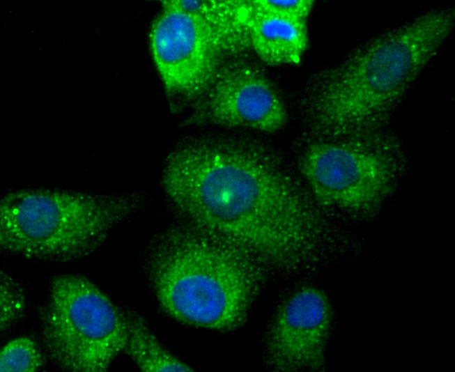
ICC staining TMS1 in A549 cells (green). The nuclear counter stain is DAPI (blue). Cells were fixed in paraformaldehyde, permeabilised with 0.25% Triton X100/PBS.
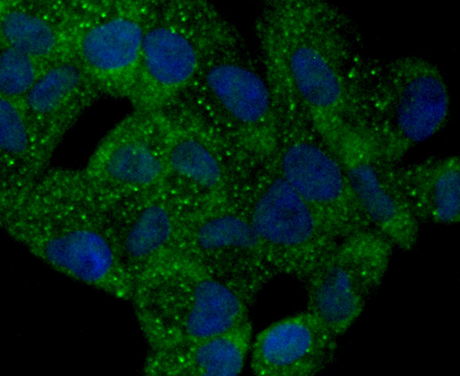
ICC staining TMS1 in Hela cells (green). The nuclear counter stain is DAPI (blue). Cells were fixed in paraformaldehyde, permeabilised with 0.25% Triton X100/PBS.
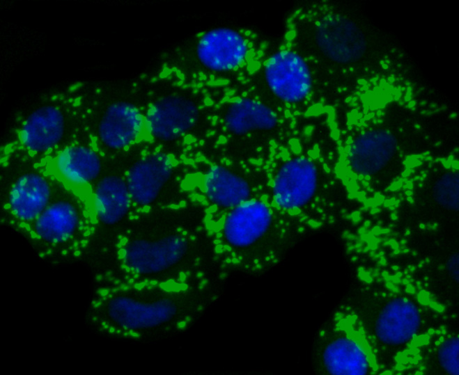
ICC staining TMS1 in HepG2 cells (green). The nuclear counter stain is DAPI (blue). Cells were fixed in paraformaldehyde, permeabilised with 0.25% Triton X100/PBS.
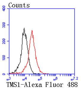
Flow cytometric analysis of 293 cells with TMS1 antibody at 1/50 dilution (red) compared with an unlabelled control (cells without incubation with primary antibody; black). Alexa Fluor 488-conjugated goat anti rabbit IgG was used as the secondary antibody
The death domain (DD) superfamily of proteins share one or more of the following domains: the DD, DED (death-effector domain), CARD (caspase-recruitment domain) and PYD (Pyrin domain). Each of these domains is characterized by a canonical death domain fold, which consists of a bundle of five or six antiparallel α-helices. As their names suggest, these domains play prominent roles in programmed cell death. Caspase-associated recruitment domains (CARDs) mediate the interaction between adaptor proteins such as Apaf-1 and the proform of caspases (e.g., CASP9) participating in apoptosis. ASC (apoptosis-associated speck-like protein containing a CARD, also known as TMS1or PYCARD) is a member of the CARD-containing adaptor protein family. ASC is a 195 amino acid protein, containing a N-terminal Pyrin-like domain (PYD) and an 87 residue C-terminal CARD. This motif is characteristic of numerous proteins involved in apoptotic signaling. ASC2 (apoptosis-associated speck-like protein containing a CARD 2), also known as Pyrin-only protein 1 or PADD-only protein 1, is an 89 amino acid member of the DD superfamily that contains one Pyrin domain. Localized to the cytoplasm, ASC2 interacts with ASC to modulate NF-κB and pro-caspase-1 regulation.
If you have published an article using product 49077, please notify us so that we can cite your literature.

 Yes
Yes