Product Detail
Product NameCytokeratin 4 Rabbit mAb
Clone No.SN74-03
Host SpeciesRecombinant Rabbit
Clonality Monoclonal
PurificationProA affinity purified
ApplicationsWB, ICC/IF, IHC, IP, FC
Species ReactivityHu
Immunogen Descrecombinant protein
ConjugateUnconjugated
Other NamesCK 4 antibody CK-4 antibody CK4 antibody CYK4 antibody Cytokeratin 4 antibody Cytokeratin-4 antibody Cytokeratin4 antibody FLJ31692 antibody K2C4_HUMAN antibody K4 antibody Keratin 4 antibody Keratin antibody Keratin type II cytoskeletal 4 antibody Keratin-4 antibody Keratin4 antibody KRT 4 antibody Krt4 antibody type II cytoskeletal 4 antibody Type-II keratin Kb4 antibody
Accession NoSwiss-Prot#:P19013
Uniprot
P19013
Calculated MW57 kDa
Formulation1*TBS (pH7.4), 1%BSA, 40%Glycerol. Preservative: 0.05% Sodium Azide.
StorageStore at -20˚C
Application Details
WB: 1:500-1:1000
IHC: 1:50-1:200
ICC: 1:50-1:200
FC: 1:50-1:100
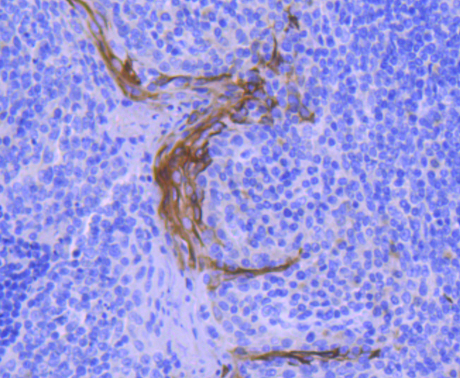
Immunohistochemical analysis of paraffin-embedded human tonsil tissue using anti-Cytokeratin 4 antibody. Counter stained with hematoxylin.
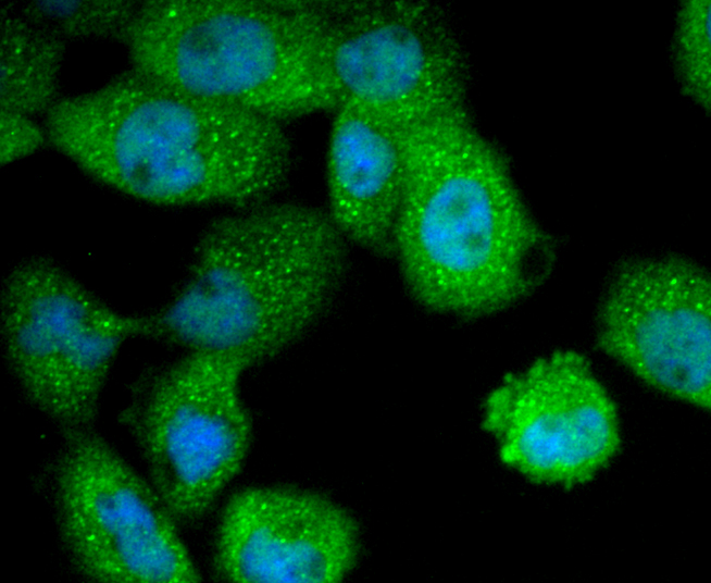
ICC staining Cytokeratin 4 in A431 cells (green). The nuclear counter stain is DAPI (blue). Cells were fixed in paraformaldehyde, permeabilised with 0.25% Triton X100/PBS.
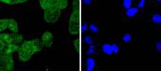
ICC staining Cytokeratin 4 in Hela cells (green). The nuclear counter stain is DAPI (blue). Cells were fixed in paraformaldehyde, permeabilised with 0.25% Triton X100/PBS.
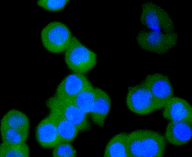
ICC staining Cytokeratin 4 in SW480 cells (green). The nuclear counter stain is DAPI (blue). Cells were fixed in paraformaldehyde, permeabilised with 0.25% Triton X100/PBS.
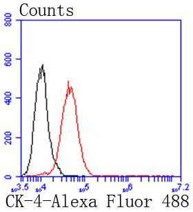
Flow cytometric analysis of A431 cells with Cytokeratin 4 antibody at 1/50 dilution (red) compared with an unlabelled control (cells without incubation with primary antibody; black). Alexa Fluor 488-conjugated goat anti rabbit IgG was used as the secondary antibody.
Cytokeratins are a subfamily of intermediate filament keratins that are characterized by a remarkable biochemical diversity, which is represented in human epithelial tissues by at least 20 different polypeptides. Cytokeratins range in isoelectric range between 4.9 and 7.8. Cytokeratin 1 has the highest molecular weight, while Cytokeratin 19 has the lowest molecular weight. The cytokeratins are divided into the type I and type II subgroups. Type II family members comprise the basic to neutral members, Cytokeratins 1-8, while the type I group comprises the acidic members, Cytokeratins 9-20. Various epithelia in the human body usually express cytokeratins which are characteristic of the type of epithelium and related to the degree of maturation or differentiation within said epithelium. Cytokeratin subtype expression patterns are used to an increasing extent in the distinction of different types of epithelial malignancies. Cytokeratin 4 is expressed in differentiated layers of the mucosal and esophageal epithelia along with Cytokeratin 13.
If you have published an article using product 49087, please notify us so that we can cite your literature.


 Yes
Yes


