Product Detail
Product NameCyclin H Rabbit mAb
Clone No.SN20-48
Host SpeciesRecombinant Rabbit
Clonality Monoclonal
PurificationProA affinity purified
ApplicationsWB, ICC/IF, IHC, IP, FC
Species ReactivityHu, Ms
Immunogen Descrecombinant protein
ConjugateUnconjugated
Other Names6330408H09Rik antibody AI661354 antibody AV102684 antibody AW538719 antibody CAK antibody CAK complex subunit antibody ccnh antibody CCNH_HUMAN antibody CDK activating kinase antibody CDK activating kinase complex subunit antibody Cyclin dependent kinase activating kinase antibody cyclin dependent kinase activating kinase complex subunit antibody Cyclin H antibody Cyclin-H antibody CyclinH antibody MO15 associated protein antibody MO15-associated protein antibody p34 antibody p36 antibody p37 antibody
Accession NoSwiss-Prot#:P51946
Uniprot
P51946
Gene ID
902;
Calculated MW38 kDa
Formulation1*TBS (pH7.4), 1%BSA, 40%Glycerol. Preservative: 0.05% Sodium Azide.
StorageStore at -20˚C
Application Details
WB: 1:1,000-5,000
IHC: 1:50-1:200
ICC: 1:100-1:500
FC: 1:50-1:100
Western blot analysis of Cyclin H on K562 cells lysates using anti-Cyclin H antibody at 1/1,000 dilution.
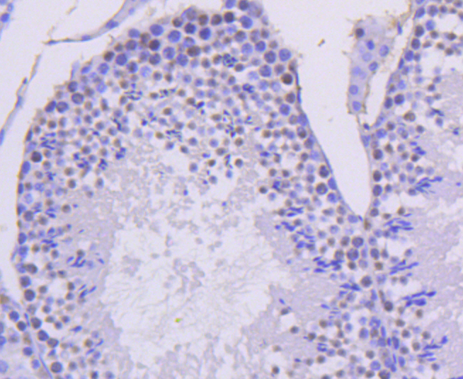
Immunohistochemical analysis of paraffin-embedded mouse testis tissue using anti-Cyclin H antibody. Counter stained with hematoxylin.
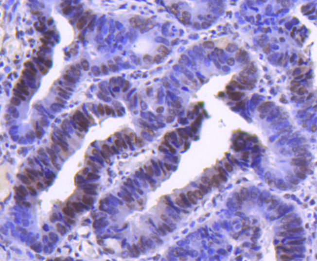
Immunohistochemical analysis of paraffin-embedded human colon cancer tissue using anti-Cyclin H antibody. Counter stained with hematoxylin.
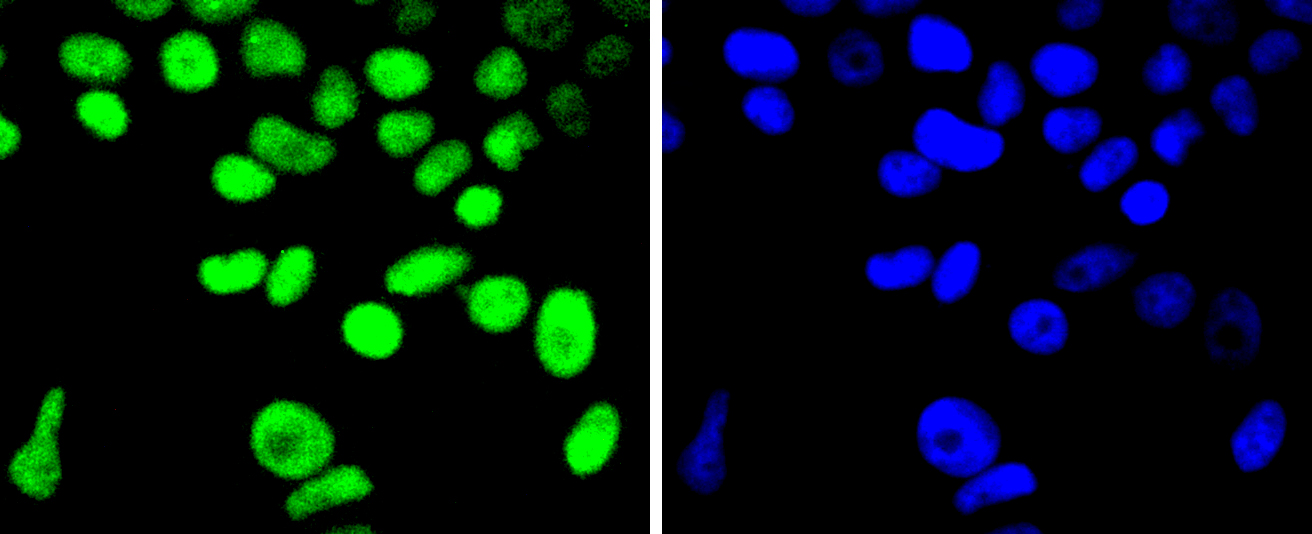
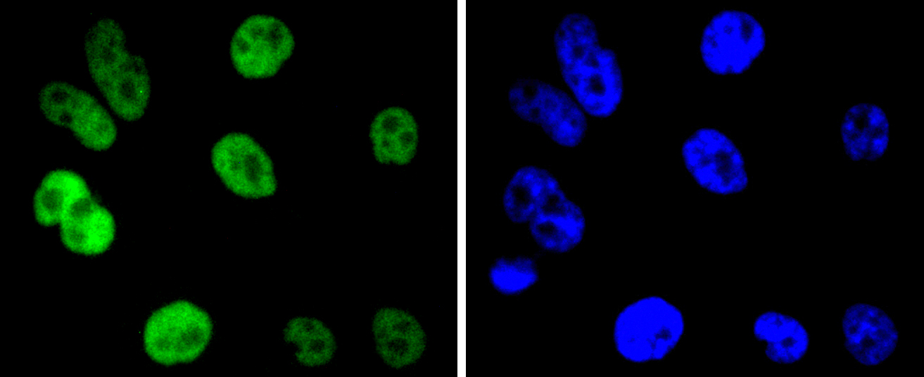
ICC staining Cyclin H in Hela cells (green). The nuclear counter stain is DAPI (blue). Cells were fixed in paraformaldehyde, permeabilised with 0.25% Triton X100/PBS.
ICC staining Cyclin H in MCF-7 cells (green). The nuclear counter stain is DAPI (blue). Cells were fixed in paraformaldehyde, permeabilised with 0.25% Triton X100/PBS.
ICC staining Cyclin H in PC-3M cells (green). The nuclear counter stain is DAPI (blue). Cells were fixed in paraformaldehyde, permeabilised with 0.25% Triton X100/PBS.
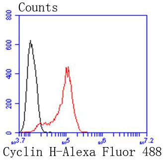
Flow cytometric analysis of Hela cells with Cyclin H antibody at 1/50 dilution (red) compared with an unlabelled control (cells without incubation with primary antibody; black). Alexa Fluor 488-conjugated goat anti rabbit IgG was used as the secondary antibody.
Progression through the cell cycle requires activation of a series of enzymes designated cyclin dependent kinases (Cdks). The monomeric catalytic subunit, Cdk2, a critical enzyme for initiation of cell cycle progression, is completely inactive. Partial activation is achieved by the binding of regulatory cyclins such as cyclin D1, while full activation requires, in addition, phosphorylation at Thr-160. The enzyme responsible for phosphorylation of Thr-160 in Cdk2 and also Thr-161 in Cdc2 p34, designated Cdk-activating kinase (CAK), has been partially purified and shown to be comprised of a catalytic subunit and a regulatory subunit. The catalytic subunit, designated Cdk7, has been identified as the mammalian homolog of MO15, a protein kinase demonstrated earlier in starfish and Xenopus. The regulatory subunit is a novel cyclin (cyclin H) and is required for activation of Cdk7. Like other Cdks, Cdk7 contains a conserved threonine required for full activity; mutation of this residue severely reduces CAK activity.
If you have published an article using product 49100, please notify us so that we can cite your literature.


 Yes
Yes


