Product Detail
Product NameGlutamate receptor 1 Rabbit mAb
Clone No.SD2010
Host SpeciesRecombinant Rabbit
Clonality Monoclonal
PurificationProA affinity purified
ApplicationsWB, IHC, IP
Species ReactivityHu, Ms, Rt
Immunogen Descrecombinant protein
ConjugateUnconjugated
Other NamesGLUR 1 antibody
GLUR A antibody
AMPA 1 antibody
AMPA selective glutamate receptor 1 antibody
AMPA-selective glutamate receptor 1 antibody
GluA1 antibody
GLUH1 antibody
GluR K1 antibody
GluR-1 antibody
GluR-A antibody
GluR-K1 antibody
GLUR1 antibody
GLURA antibody
GluRK1 antibody
Glutamate receptor 1 antibody
Glutamate receptor ionotropic AMPA 1 antibody
Glutamate receptor ionotropic antibody
Glutamate receptor, ionotropic, AMPA 1 antibody
Gria1 antibody
GRIA1_HUMAN antibody
HBGR1 antibody
MGC133252 antibody
OTTHUMP00000160643 antibody
OTTHUMP00000165781 antibody
OTTHUMP00000224241 antibody
OTTHUMP00000224242 antibody
OTTHUMP00000224243 antibody
Accession NoSwiss-Prot#:P42261
Uniprot
P42261
Gene ID
2890;
Calculated MW92 kDa
Formulation1*TBS (pH7.4), 1%BSA, 40%Glycerol. Preservative: 0.05% Sodium Azide.
StorageStore at -20˚C
Application Details
WB: 1:1,000-1:2,000
IHC: 1:50-1:200
Western blot analysis of GluR1 on rat brain lysates using anti-GluR1 antibody at 1/1,000 dilution.
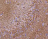
Immunohistochemical analysis of paraffin-embedded rat brain tissue using anti-GluR1 antibody. Counter stained with hematoxylin.
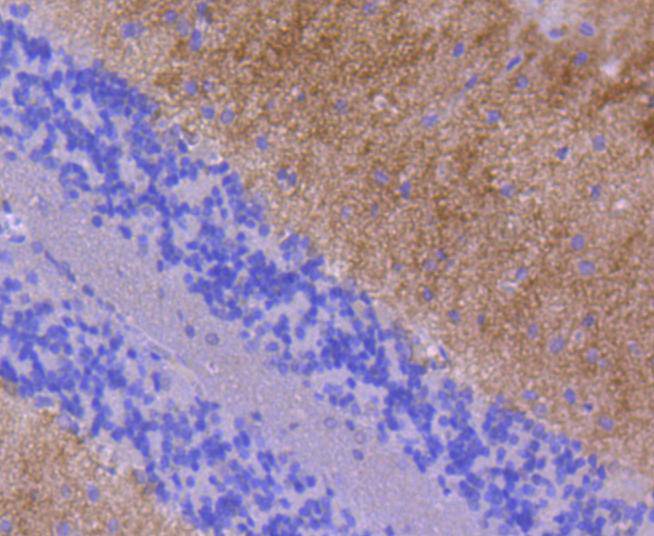
Immunohistochemical analysis of paraffin-embedded rat cerebellum tissue using anti-GluR1 antibody. Counter stained with hematoxylin.
Immunohistochemical analysis of paraffin-embedded mouse brain tissue using anti-GluR1 antibody. Counter stained with hematoxylin.
Immunohistochemical analysis of paraffin-embedded mouse cerebellum tissue using anti-GluR1 antibody. Counter stained with hematoxylin.
Glutamate receptors mediate most excitatory neurotransmission in the brain and play an important role in neural plasticity, neural development and neurodegeneration. Ionotropic glutamate receptors are categorized into NMDA receptors and kainate/AMPA receptors, both of which contain glutamate-gated, cation-specific ion channels. Kainate/AMPA receptors are co-localized with NMDA receptors in many synapses and consist of seven structurally related subunits designated GluR-1 to -7. The kainate/AMPA receptors are primarily responsible for the fast excitatory neuro-transmission by glutamate whereas the NMDA receptors are functionally characterized by a slow kinetic and a high permeability for Ca2+ ions. The NMDA receptors consist of five subunits: epsilion 1, 2, 3, 4 and one zeta subunit. The zeta subunit is expressed throughout the brainstem whereas the four epsilon subunits display limited distribution.
If you have published an article using product 49118, please notify us so that we can cite your literature.


 Yes
Yes


