Product Detail
Product NameHistone H2B Rabbit mAb
Clone No.SD20-63
Host SpeciesRecombinant Rabbit
Clonality Monoclonal
PurificationProA affinity purified
ApplicationsWB, ICC/IF, IHC, IP, FC
Species ReactivityHu, Ms, Rt
Immunogen Descrecombinant protein
ConjugateUnconjugated
Other NamesH2B GL105 antibody H2B histone family member O antibody H2B histone family member S antibody H2B.1 antibody H2B.1 B antibody H2B.b antibody H2B.c antibody H2B.d antibody H2B.e antibody H2B.f antibody H2B.j antibody H2B.q antibody H2B/b antibody H2B/c antibody H2B/d antibody H2B/e antibody H2B/f antibody H2B/j antibody H2B/o antibody H2B/q antibody H2BFB antibody H2BFC antibody H2BFD antibody H2BFE antibody H2BFF antibody H2BFJ antibody H2BFO antibody H2BFQ antibody H2BFS antibody HIRIP2 antibody HIST1H2BB antibody HIST1H2BD antibody HIST1H2BH antibody HIST1H2BL antibody HIST1H2BM antibody HIST1H2BN antibody HIST2H2BE antibody Histone H2B antibody Histone H2B type 1 B antibody Histone H2B type 1 D antibody Histone H2B type 1 H antibody Histone H2B type 1 L antibody Histone H2B type 1 M antibody Histone H2B type 1 N antibody Histone H2B type 2 E antibody histone protein antibody
Accession NoSwiss-Prot#:O60814
Uniprot
O60814
Gene ID
85236;
Calculated MW14 kDa
Formulation1*TBS (pH7.4), 1%BSA, 40%Glycerol. Preservative: 0.05% Sodium Azide.
StorageStore at -20˚C
Application Details
WB: 1:1,000-5,000
IHC: 1:50-1:200
ICC: 1:100-1:500
FC: 1:50-1:100
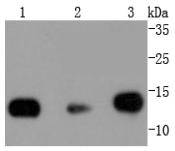
Western blot analysis of Histone H2B on different lysates using anti-Histone H2B antibody at 1/1,000 dilution. Positive control: Lane 1: Hela Lane 2: NIH/3T3 Lane 3: PC12
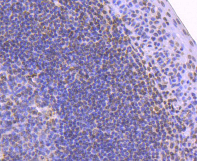
Immunohistochemical analysis of paraffin-embedded human tonsil tissue using anti-Histone H2B antibody. Counter stained with hematoxylin.
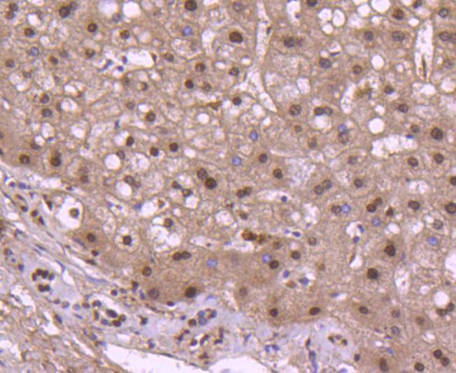
Immunohistochemical analysis of paraffin-embedded human liver tissue using anti-Histone H2B antibody. Counter stained with hematoxylin.
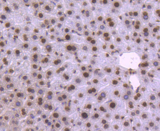
Immunohistochemical analysis of paraffin-embedded mouse liver tissue using anti-Histone H2B antibody. Counter stained with hematoxylin.
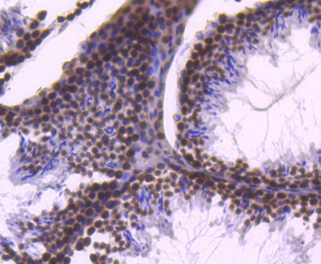
Immunohistochemical analysis of paraffin-embedded mouse testis tissue using anti-Histone H2B antibody. Counter stained with hematoxylin.
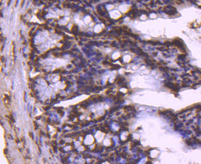
Immunohistochemical analysis of paraffin-embedded mouse colon tissue using anti-Histone H2B antibody. Counter stained with hematoxylin.
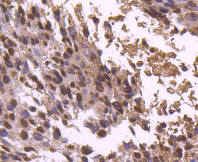
Immunohistochemical analysis of paraffin-embedded human breast carcinoma tissue using anti-Histone H2B antibody. Counter stained with hematoxylin.
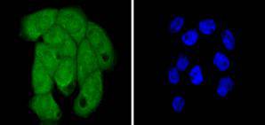
ICC staining Histone H2B in A431 cells (green). The nuclear counter stain is DAPI (blue). Cells were fixed in paraformaldehyde, permeabilised with 0.25% Triton X100/PBS.
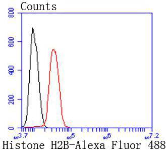
Flow cytometric analysis of Hela cells with Histone H2B antibody at 1/50 dilution (red) compared with an unlabelled control (cells without incubation with primary antibody; black). Alexa Fluor 488-conjugated goat anti rabbit IgG was used as the secondary antibody.
Eukaryotic histones are basic and water soluble nuclear proteins that form hetero-octameric nucleosome particles by wrapping 146 base pairs of DNA in a left-handed super-helical turn sequentially to form chromosomal fiber. Two molecules of each of the four core histones (H2A, H2B, H3, and H4) form the octamer; formed of two H2A-H2B dimers and two H3-H4 dimers, forming two nearly symmetrical halves by tertiary structure. Over 80% of nucleosomes contain the linker Histone H1, derived from an intronless gene, that interacts with linker DNA between nucleosomes and mediates compaction into higher order chromatin. Histones are subject to posttranslational modification by enzymes primarily on their N-terminal tails, but also in their globular domains. Such modifications include methylation, citrullination, acetylation, phosphorylation, sumoylation, ubiquitination and ADP-ribosylation.
If you have published an article using product 49134, please notify us so that we can cite your literature.


 Yes
Yes


