Welcome to Sabbiotech!     
Email:info@signalwayantibody.com
Location: Home > Recombinant Rabbit Monoclonal Antibodies > CD36 Rabbit mAb

CD36 Rabbit mAb#49232

Availability: Yes
Total Amount:$
Product Detail

Product NameCD36 Rabbit mAb

Clone No.SR4532

Host SpeciesRecombinant Rabbit

ClonalityMonoclonal

PurificationProA affinity purified

ApplicationsWB, ICC/IF, IHC, IP, FC

Species ReactivityHu, Ms, Rt

Immunogen Descrecombinant protein

ConjugateUnconjugated

Other NamesAdipocyte membrane protein antibody
BDPLT10 antibody
CD36 antibody
CD36 antigen (collagen type I receptor, thrombospondin receptor) antibody
CD36 antigen antibody
CD36 molecule (thrombospondin receptor) antibody
CD36 molecule antibody
CD36_HUMAN antibody
CHDS7 antibody
Cluster determinant 36 antibody
Collagen receptor, platelet antibody
FAT antibody
Fatty acid translocase antibody
Fatty acid transport protein antibody
Glycoprotein IIIb antibody
GP IIIb antibody
GP3B antibody
GP4 antibody
GPIIIB antibody
GPIV antibody
Leukocyte differentiation antigen CD36 antibody
MGC108510 antibody
MGC91634 antibody
PAS 4 protein antibody
PAS IV antibody
PAS-4 antibody
PASIV antibody
Platelet collagen receptor antibody
Platelet glycoprotein 4 antibody
Platelet glycoprotein IV antibody
scarb3 antibody
Scavenger receptor class B member 3 antibody
Thrombospondin receptor antibody

Accession NoSwiss-Prot#:P16671

Uniprot P16671

Gene ID 948;

Calculated MW53 kDa

Formulation1*TBS (pH7.4), 1%BSA, 40%Glycerol. Preservative: 0.05% Sodium Azide.

StorageStore at -20˚C

Application Details
WB: 1:1,000
IHC: 1:50-1:200
ICC: 1:50-1:200

FC: 1:50-1:100
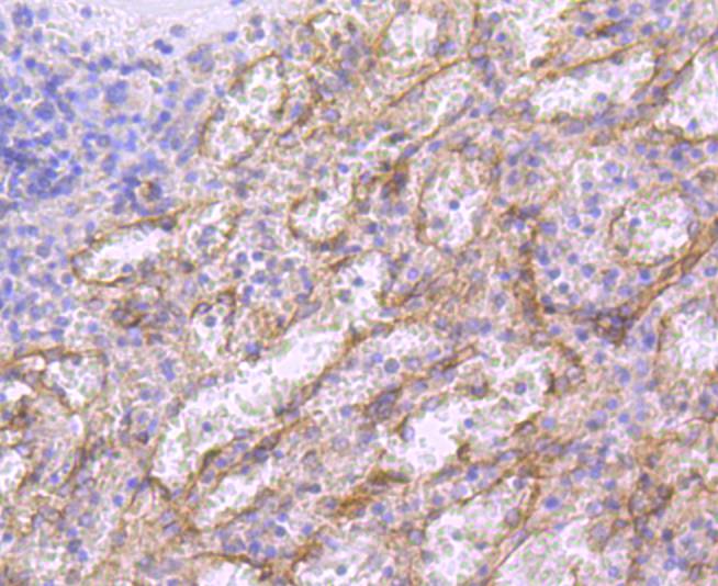
Immunohistochemical analysis of paraffin-embedded human spleen tissue using anti-CD36 antibody. Counter stained with hematoxylin.
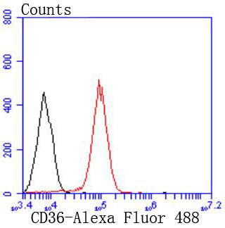
Flow cytometric analysis of THP-1 cells with CD36 antibody at 1/50 dilution (red) compared with an unlabelled control (cells without incubation with primary antibody; black). Alexa Fluor 488-conjugated goat anti rabbit IgG was used as the secondary antibody.
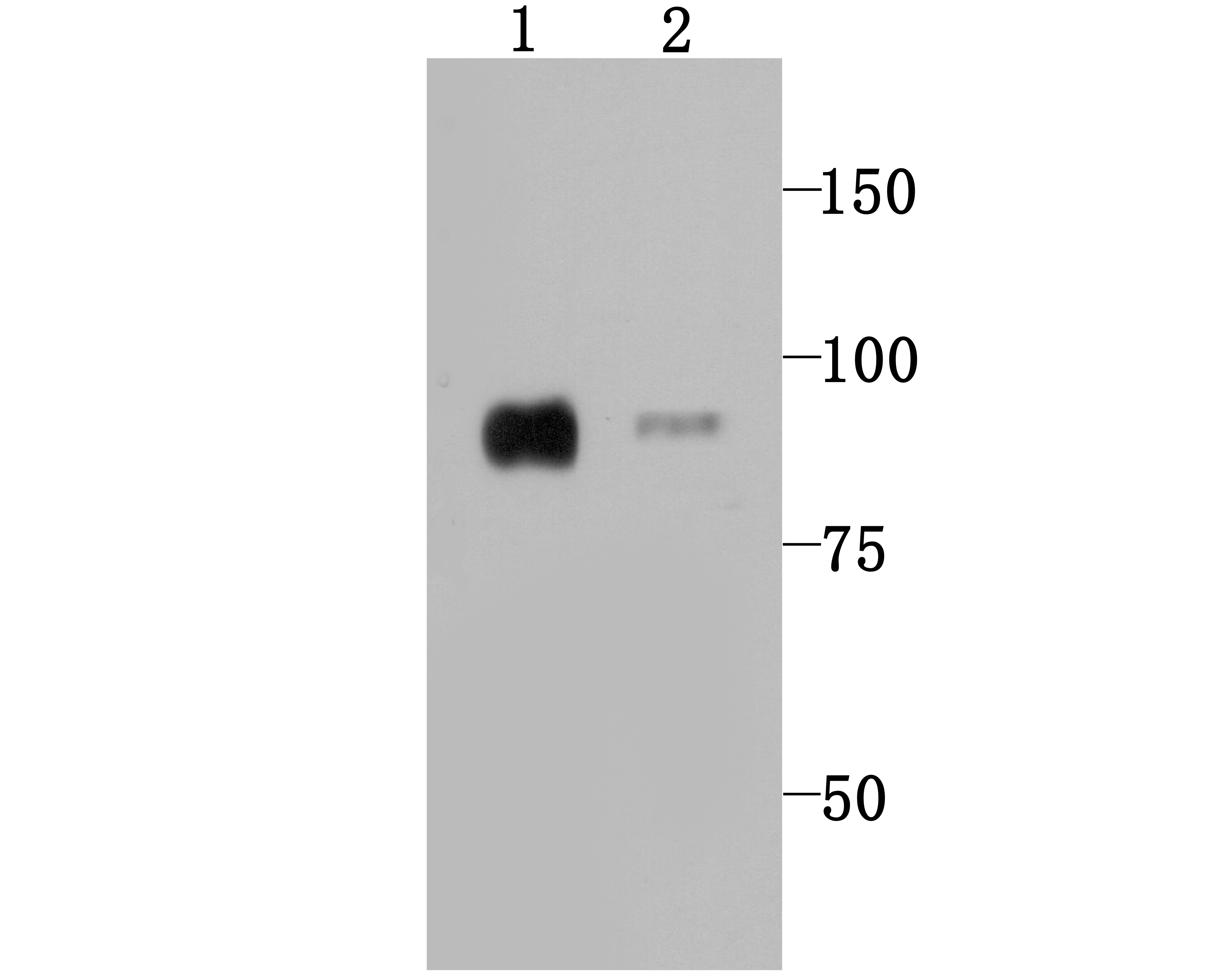
Western blot analysis of CD36 on human heart (1) and mouse heart (2) tissue lysates using anti-CD36 antibody at 1/1,000 dilution.
ICC staining CD36 in SH-SY5Y cells (green). The nuclear counter stain is DAPI (blue). Cells were fixed in paraformaldehyde, permeabilised with 0.25% Triton X100/PBS.
CD36 (collagen type I receptor, thrombospondin receptor, FAT, GP4, GP3B, GPIV, PASIV, SCARB3) is a membrane glycoprotein on platelets, monocytes and umbilical vein endothelial cells. CD36 binds to collagen, thrombospondin, anionic phospholipids and oxidized LDL. CD36 plays a key role in both phagocytosis and lipid recycling, for constant production of mature spermatozoa. Mutations in this gene cause platelet glycoprotein deficiency. Three alternatively spliced transcript variants encoding the same protein isoform have been found for this gene. Thrombospondins are widely distributed proteins that influence a variety of adhesive processes and CD36 may have important functions as a cell adhesion molecule.

If you have published an article using product 49232, please notify us so that we can cite your literature.

NOTE

Application

  • WBWestern Blotting
  • IHCImmunohistochemistry
  • IFImmunofluorescence
  • ICCImmunocytochemistry
  • FCFlow Cytometry
  • IPImmunoprecipitation
  • EELISA
  • DBDot Blotting
  • ChIPChromatin Immunoprecipitation
  • GICAGold Immunochromatography Assay
  • NCNegative Control

Species Reactivity

  • HuHuman
  • MsMouse
  • RtRat
  • DmDrosophila melanogaster
  • CCaenorhabditis elegans
  • MkMonkey
  • RbRabbit
  • BBovine
  • DDog
  • PPig
  • HmHamster
  • ChHmChinese Hamster
  • ChkChicken
  • ShpSheep
;