Product Detail
Product NameFUS/TLS Rabbit mAb
Clone No.JJ09-31
Host SpeciesRecombinant Rabbit
Clonality Monoclonal
PurificationProA affinity purified
ApplicationsWB, ICC/IF, IHC, FC
Species ReactivityHu, Ms, Rt
Immunogen Descrecombinant protein
ConjugateUnconjugated
Other Names75 kDa DNA pairing protein antibody
75 kDa DNA-pairing protein antibody
ALS6 antibody
Amyotrophic lateral sclerosis 6 antibody
fus antibody
FUS CHOP antibody
Fus like protein antibody
FUS_HUMAN antibody
FUS1 antibody
Fused in sarcoma antibody
Fusion (involved in t(12;16) in malignant liposarcoma) antibody
Fusion derived from t(12;16) malignant liposarcoma antibody
Fusion gene in myxoid liposarcoma antibody
Heterogeneous nuclear ribonucleoprotein P2 antibody
hnRNP P2 antibody
hnRNPP2 antibody
Oncogene FUS antibody
Oncogene TLS antibody
POMp75 antibody
RNA binding protein FUS antibody
RNA-binding protein FUS antibody
TLS antibody
TLS CHOP antibody
Translocated in liposarcoma antibody
Translocated in liposarcoma protein antibody
Accession NoSwiss-Prot#:P35637
Uniprot
P35637
Gene ID
2521;
Calculated MW75 kDa
Formulation1*TBS (pH7.4), 1%BSA, 40%Glycerol. Preservative: 0.05% Sodium Azide.
StorageStore at -20˚C
Application Details
WB: 1:1,000
IHC: 1:50-1:200
ICC: 1:50-1:200
FC: 1:50-1:100
Western blot analysis of FUS/TLS on K562 cells lysates using anti-FUS/TLS antibody at 1/1,000 dilution.
Immunohistochemical analysis of paraffin-embedded human kidney tissue using anti-FUS/TLS antibody. Counter stained with hematoxylin.
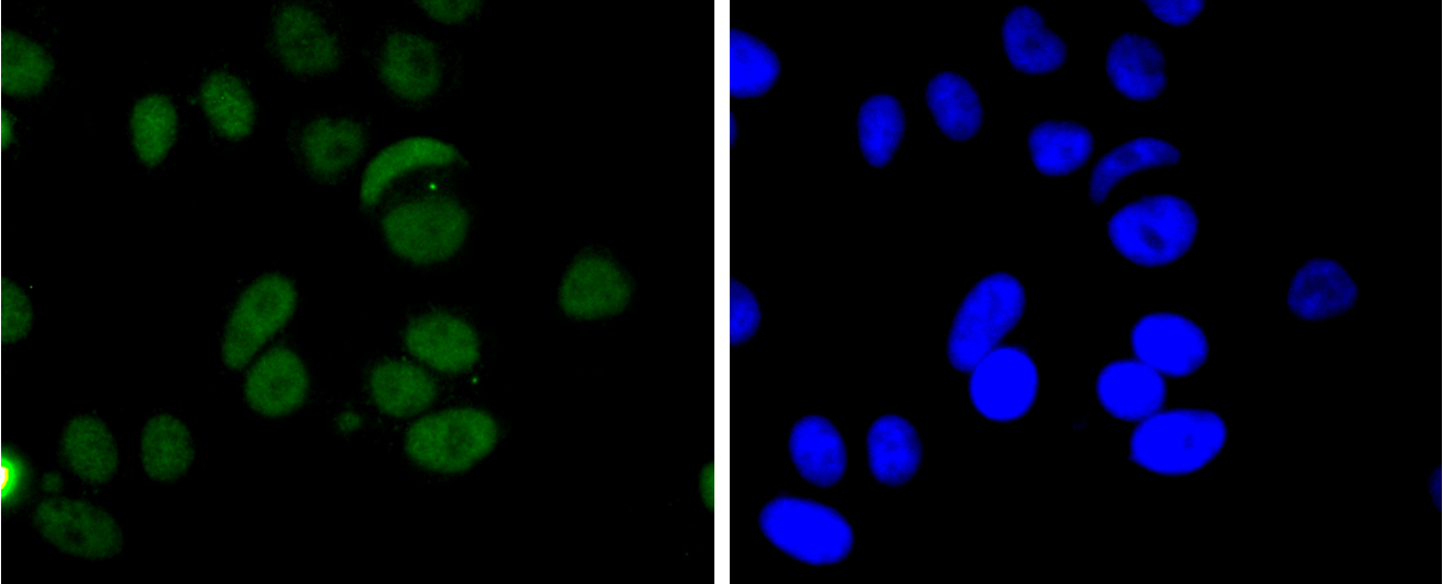
ICC staining FUS/TLS in Hela cells (green). The nuclear counter stain is DAPI (blue). Cells were fixed in paraformaldehyde, permeabilised with 0.25% Triton X100/PBS.
ICC staining FUS/TLS in MCF-7 cells (green). The nuclear counter stain is DAPI (blue). Cells were fixed in paraformaldehyde, permeabilised with 0.25% Triton X100/PBS.
ICC staining FUS/TLS in SW480 cells (green). The nuclear counter stain is DAPI (blue). Cells were fixed in paraformaldehyde, permeabilised with 0.25% Triton X100/PBS.
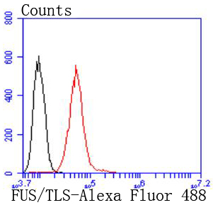
Flow cytometric analysis of Hela cells with FUS/TLS antibody at 1/50 dilution (red) compared with an unlabelled control (cells without incubation with primary antibody; black). Alexa Fluor 488-conjugated goat anti rabbit IgG was used as the secondary antibody.
EWS and FUS/TLS are nuclear RNA-binding proteins. As a result of chromosome translocation, the EWS gene is fused to a variety of transcription factors, including ATF-1, in human neoplasias. In the Ewing family of tumors, the N-terminal domain of EWS is fused to the DNA-binding domain of various Ets transcription factors, including Fli-1, ETV1 and FEV. The EWS/Fli-1 chimeric protein acts as a more potent transcriptional activator than Fli-1 and can promote cell transformation. In human myxoid liposarcomas and myeloid leukemias, chromosomal translocation results in the fusion of the N-terminal region of FUS/TLS with the open reading frame of CHOP. In normal cells, FUS/TLS binds to the DNA-binding domains of nuclear steroid receptors and is also present in subpopulations of TFIID complexes, indicating a potential role for FUS/TLS in the processing of primary transcripts that are generated in response to hormone-induced transcription.
If you have published an article using product 49300, please notify us so that we can cite your literature.


 Yes
Yes


